TMIP
Our Vision
To advance the timely translation of basic molecular imaging research to clinical environments by connecting researchers and clinicians across Texas institutions and nationally.
Our Mission
To facilitate translation of novel molecular imaging radiopharmaceuticals and targeted molecular radiotheragnostics to patient care enabled by our integrated network of highly skilled physicians and scientists.
About Us
We work closely with the department's Cyclotron and Radiochemistry Program to commission locally synthesized molecular diagnostics and therapeutics. The on-site Cyclotron suite has uniquely positioned the TMIP to aid with molecular imaging projects at any stage across the translational research continuum. We are equipped with the ability to translate a diagnostic or therapeutic agent from it's in silico inception to in vitro validation studies, on to biodistribution, safety, and efficacy studies in preclinical models, and finally to the clinical trials that directly serve our patients.
- Faculty & Staff
Faculty

Professor
Program Director, Nuclear Medicine Residency
Research Lab
Assistant Professor
Affiliated Faculty
- Research Projects
Research Projects
- Associated Cores
Associated Cores
DASPA
The Data Storage, Processing & Analysis core provides informatics support to researchers in a variety of areas.
Research PACS
- iPACS provides a HIPAA-compliant PACS system for archiving clinical and preclinical research imaging studies
- The installed and supported iPACS system is a web-based, secure, project-oriented resource available to Faculty researchers in Radiology and their collaborators
- Also has the capability of performing customized de-identification of images acquired in clinical trials to preserve patient confidentiality
ANSIR
The Advanced NeuroScience Imaging Research (ANSIR) lab dataset processing provides:
- Fully automated analysis of neuro MRI data including structural analysis using SPM and Freesurfer, task and resting state fMRI processing, Diffusion Tensor image processing, automated FLAIR white matter lesion segmentation, Arterial Spin Label MRI processing, and quantitative susceptibility mapping.
- Project-specific XNAT database storage allowing retrieval and queries of imaging and metadata
- Clinical alerts without a formal report can be provided for incidental findings
- Additional custom project-specific services may be available following consultation and based on resource availability
IM4T
The UTSW Radiology Department's Imaging Metrics for Trials (IM4T) group provides diagnostic imaging interpretation of cancer treatment response evaluation to internal and external researchers involved in cancer research studies that require response evaluation using Response Evaluation Criteria in Solid Tumors (RECIST) and its variants.
Magnetic Resonance Imaging (MRI)
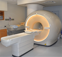
The Magnetic Resonance (MR) core is established to facilitate research and development within the Department of Radiology and collaborating departments in the field of MR imaging (MRI) and MR spectroscopy (MRS). The MR core consists of a whole-body human scanner (Philips Ingenia 3T MR) and a small animal Desktop scanner (Aspect 1T MR). Philips Ingenia includes dual-transmit and digital architecture for signal reception. This scanner enables the development and evaluation of new MRI/MRS techniques for improved diagnosis and understanding of the pathophysiology of disease. Aspect 1T scanner enables sequential imaging of small animals (e.g. mice and rats), without sacrificing them, for longitudinal monitoring of disease progression and/or therapy response. Being cited next to the small animal PET/CT and SPECT/CT scanners, this allows superposition of images for multimodality analysis.
Click here to explore more about the MR Core.
Translational Molecular Imaging Core (TMIC)
- Cyclotron and radiochemistry facility approved for CGMP production of PET radiopharmaceuticals for human use. Capable of producing 6 radioisotopes and >30 radiotracers in addition to the FDA-approved tracers
- A regulatory office in the Department of Radiology facilitates Investigational New Drug (IND) and Abbreviated New Drug Applications (ANDA) approval of radiotracers.
- Radiochemistry and nuclear medicine experts to advise investigators on the development and implementation of imaging protocols in a range of disease models (e.g., cancer, diabetes, metabolism, cardiotoxicity, neurodegenerative diseases, etc.)
- Pre-clinical imaging including a Siemens Inveon PET/CT scanner for small animal imaging
- State-of-the-art human imaging in the NE2 building including a GE Discovery IQ five ring PET/CT scanner and a Siemens 3T Biograph hybrid PET/MR scanner, both located in close proximity to our cyclotron facility
- Publications
Publications
Faculty
Affiliated Faculty