CACTUS
Our Vision
Ultrasound is an impactful, cost-effective, and versatile tool in diagnostic imaging and intervention, and holds great promise in areas of molecular imaging and theranostics. With dedication and ingenuity our programs and people aim to realize these benefits.
Our Mission
To advance the use of ultrasound in diagnostic imaging and image-guided therapies through education, quality improvement, and clinical and translational research to optimize patient outcomes.
About Us
Who We Are
The Collaborative for Advanced Clinical Techniques in UltraSound (CACTUS) constitutes a group of like-minded physicians, scientists, and technical experts dedicated to the advancement of clinical ultrasound imaging and image-guided intervention.
What We Do
CACTUS is at the intersection of high-quality clinical care, emerging and cutting-edge clinical techniques, and translational research.
- Clinical Care - Through a robust quality-assurance program, evidence-based literature, and societal guidelines, new clinical imaging protocols are developed and tested. The impact on cost-effectiveness, workflow, and patient management are analyzed. These protocols and recommendations are then disseminated throughout the UT system and presented at local and national meetings to help impact best-practice medicine.
- New Techniques - CACTUS is focused on vetting new ultrasound-based technologies before they may be widely available. Through collaborations with manufacturers and other third party partners, CACTUS offers the expertise, personnel, and facilities to test emerging technologies to help assess their performance in a clinical setting, and their impact on patient management.
- Translational Research - CACTUS also provides the environment and infrastructure to test emerging techniques and technologies that may have been analyzed in the lab, but have not yet been performed in a clinical situation. This translation (bench-to-bedside) epitomizes the impact that CACTUS strives to achieve.
- Faculty & Staff
Affiliated Clinical Experts
Assistant Professor
Medical Director, Ultrasound
Assistant Professor
Professor
Medical Director, Genitourinary Imaging, Parkland Hospital
Professor
Affiliated Research Experts
Assistant Professor
Assistant Professor
Research Lab
Assistant Professor
Research Lab
Professor
Director, Ultrasound Research
Executive Academic Mentor
Research Lab - Research Projects
Research Projects
Highlights include,
- Development and testing of new and enhanced ultrasound techniques in a clinical setting
- Retrospective and prospective analysis of imaging and clinical data related to abdominal and pelvic ultrasound examinations
Clinical Research
- Chronic Liver Disease
- Screening and outcomes in patients with hepatocellular carcinoma (HCC)
- Fat and fibrosis quantification
- Head and Neck Cancer
- Thyroid cancer imaging
- Cervical lymph node evaluation
Translational Research Projects
- Liver fat, inflammation, and fibrosis quantification in non-alcoholic fatty liver disease (NAFLD) and non-alcoholic steatohepatitis (NASH)
- Detection of hydrogen peroxide in abscess fluid
- Use of strain and shear wave elastography in superficial ultrasound
- Quantitative contrast-enhanced ultrasound for organ and tumor perfusion analysis
- Contrast-enhanced ultrasound motion correction and frame filtering
- Ultrasound lymphangiography
Many projects are performed in collaboration with TRUST (Translational Research in UltraSound Theranostics)
- Associated Cores
Associated Cores
DASPA
The Data Storage, Processing & Analysis core provides informatics support to researchers in a variety of areas.
Research PACS
- iPACS provides a HIPAA-compliant PACS system for archiving clinical and preclinical research imaging studies
- The installed and supported iPACS system is a web-based, secure, project-oriented resource available to Faculty researchers in Radiology and their collaborators
- Also has the capability of performing customized de-identification of images acquired in clinical trials to preserve patient confidentiality
ANSIR
The Advanced NeuroScience Imaging Research (ANSIR) lab dataset processing provides:
- Fully automated analysis of neuro MRI data including structural analysis using SPM and Freesurfer, task and resting state fMRI processing, Diffusion Tensor image processing, automated FLAIR white matter lesion segmentation, Arterial Spin Label MRI processing, and quantitative susceptibility mapping.
- Project-specific XNAT database storage allowing retrieval and queries of imaging and metadata
- Clinical alerts without a formal report can be provided for incidental findings
- Additional custom project-specific services may be available following consultation and based on resource availability
IM4T
The UTSW Radiology Department's Imaging Metrics for Trials (IM4T) group provides diagnostic imaging interpretation of cancer treatment response evaluation to internal and external researchers involved in cancer research studies that require response evaluation using Response Evaluation Criteria in Solid Tumors (RECIST) and its variants.
Magnetoencephalography (MEG)
- State-of-the-art technology mapping brain function
- Most advanced MEG technology currently available, and the only MEG scanner in Dallas
- Peripheral equipment available for time-locked stimuli and responses (ear buds, button pad, accelerometers, etc.)
- Used to study various neurological disorders and injuries including dementia, autism, concussion, and many others
Magnetic Resonance Imaging (MRI)
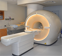
The Magnetic Resonance (MR) core is established to facilitate research and development within the Department of Radiology and collaborating departments in the field of MR imaging (MRI) and MR spectroscopy (MRS). The MR core consists of a whole-body human scanner (Philips Ingenia 3T MR) and a small animal Desktop scanner (Aspect 1T MR). Philips Ingenia includes dual-transmit and digital architecture for signal reception. This scanner enables the development and evaluation of new MRI/MRS techniques for improved diagnosis and understanding of the pathophysiology of disease. Aspect 1T scanner enables sequential imaging of small animals (e.g. mice and rats), without sacrificing them, for longitudinal monitoring of disease progression and/or therapy response. Being cited next to the small animal PET/CT and SPECT/CT scanners, this allows superposition of images for multimodality analysis.
Click here to explore more about the MR Core.
Small Animal Imaging Resource (SAIR)
- SAIR is an institutional facility that promotes and facilitates small animal imaging related to models of human disease with state-of-the-art equipment including depth resolved or planar optical imaging (fluorescence (FLI), bioluminescence (BLI) and chemiluminescence (CLI)), MRI, ultrasound, photoacoustic tomography, PET/CT, SPECT/CT and planar scintigraphy
- Infrastructure for animal handling (e.g., anesthesia, infusion, monitoring vital signs)
- Experienced investigators and technical staff capable of undertaking imaging and assisting in data interpretation are associated with the Resource and provide consultation on experimental planning, analysis and validation, and data archiving.
- Expertise in pulse programming and implementation for novel MRI experiments, design and acquisition or building of MR coils, choice of reporter molecules and /or genes, radiolabeling procedures and synthesis of ligands.
- Currently administered jointly by the Advanced Imaging Research Center (AIRC) and the Department of Radiology
Translational Molecular Imaging Core (TMIC)
- Cyclotron and radiochemistry facility approved for CGMP production of PET radiopharmaceuticals for human use. Capable of producing 6 radioisotopes and >30 radiotracers in addition to the FDA-approved tracers
- A regulatory office in the Department of Radiology facilitates Investigational New Drug (IND) and Abbreviated New Drug Applications (ANDA) approval of radiotracers.
- Radiochemistry and nuclear medicine experts to advise investigators on the development and implementation of imaging protocols in a range of disease models (e.g., cancer, diabetes, metabolism, cardiotoxicity, neurodegenerative diseases, etc.)
- Pre-clinical imaging including a Siemens Inveon PET/CT scanner for small animal imaging
- State-of-the-art human imaging in the NE2 building including a GE Discovery IQ five ring PET/CT scanner and a Siemens 3T Biograph hybrid PET/MR scanner, both located in close proximity to our cyclotron facility
- Publications
Publications
Peer-Reviewed Manuscripts
- Malone CD, Fetzer DT, Monsky WL, Itani M, Mellnick VM, Velez PA, Middleton WD, Averkiou MA, Ramaswamy RS. Contrast-enhanced US for the Interventional Radiologist: Current and Emerging Applications. RadioGraphics, 40 (2), 562- 588 Mar-Apr 2020. PMID: 32125955. DOI: 10.1148/rg.2020190183
- Choi HH, Rodgers SK, Fetzer DT, Wasnik AP, et al. Ultrasound Liver Imaging Reporting and Data System (US LI-RADS): An Overview with Technical and Practical Applications. Acad Radiol, online July 24, 2020. https://doi.org/10.1016/j.acra.2020.06.004
- Sheth S, Fetzer DT, Frates M, Needleman L, Middleton W, Jones J, Podrasky A, Gankowski-Gettle L. Guidelines for Ultrasound in the Radiology Department During the COVID-19 Pandemic. Ultrasound Quarterly, Sept 2020, 36(3): 200- 205. doi: 10.1097/RUQ.0000000000000526
- Vij A, Fetzer DT. Persistent Heterogeneous Liver Enhancement after US Contrast Material Injection. Radiology. 2019 Sep;292(3):539. PMID: 31335260 doi: 10.1148/radiol.2019190229
- Kono K, Sirlin CV, Fetzer DT, Kim TK, Rodgers SK, Piscagliall F, Lyshchik A, Dietrich CF, Wilson SR. Time to Clarify Common Misconceptions about the Liver Imaging Reporting and Data System for Contrast-enhanced US
- Fetzer DT, Flanagan J, Nabhan A, Pongsatiawong K, Antonelli J, Pearle M, Vijay K, Watumull L. Impact of Implementing Contrast‐Enhanced Ultrasound for Antegrade Nephrostogram After Percutaneous Nephrolithotomy. J Ultrasound in Medicine. July 8, 2020. https://doi.org/10.1002/jum.15380
- Elsayes KM, Kielar AZ, Chernyak V, Morshid A, Furlan A, Masch WR, Marks RM, Kamaya A, Do RKG, Kono Y, Fowler KJ, Tang A, Bashir MR, Hecht EM, Jambhekar K, Lyshchik A, Rodgers SK, Heiken JP, Kohli M, Fetzer DT, Wilson SR, Kassam Z, Mendiratta-Lala M, Singal AG, Lim CS, Cruite I, Lee J, Ash R, Mitchell DG, McInnes MDF, Sirlin CB. LI-RADS: a conceptual and historical review from its beginning to its recent integration into AASLD clinical practice guidance. J Hepatocell Carcinoma. 2019 Feb 2019;6:49-69. DOI https://doi.org/10.2147/JHC.S186239
- Rodgers SK, Fetzer DT, Gabriel H, Seow JH, Choi HH, Maturen KE, Wasnik AP, Morgan TA, Dahiya N, O'Boyle MK, Kono Y, Sirlin CB, Kamaya A. Role of US LI-RADS in the LI-RADS Algorithm. RadioGraphics. 2019;39(3):690-708. Impact Factor: 3.249. PMID: 31059393. doi: 10.1148/rg.2019180158
- Burrowes DP, Choi HH, Rodgers SK, Fetzer DT, Kamaya A. Utility of ultrasound in acute pancreatitis. Abdom Radiol (2019). Online 16 December 2019. https://doi.org/10.1007/s00261-019-02364-x
- Venkataraman V, Browning T, Pedrosa I, Abbara S, Fetzer DT, Toomay S, Peshock RM. Implementing Shared, Standardized Imaging Protocols to Improve Cross-Enterprise Workflow and Quality. 2019 Oct;32(5):880-887. PMID: 30756266 doi: 10.1007/s10278-019-00185-4
- Gleason A, Bishop K, Xi Y, Fetzer DT. Isolated Right-Sided Varicocele: Is Further Workup Necessary? Am J Roentgenol. 2019 Feb;212:1-6. PMID: 30779666. DOI: 10.2214/AJR.18.20077
- Malone CD, Fetzer DT, Lux J, Mattrey RF. Catalase-Containing Silica Particles as Ultrasound-Based Hydrogen Peroxide Sensors to Determine Infected From Noninfected Fluid Collections in Humans. AJR Am J Roentgenol. 2019 Mar 12:W1-W8. doi: 10.2214/AJR.18.20779
- Fetzer DT, Rafailidis V, Peterson C, Grant EG, Sidhu P, Barr RG. Artifacts in Contrast-Enhanced Ultrasound: a Pictorial Essay. Abdom Radiol (NY). 2018 Apr;43(4):977-997. PMID: 29198008. doi: 10.1007/s00261-017-1417-8
- Ranganath PG, Robbin ML, Back SJ, Grant EG, Fetzer DT. Practical advantages of contrast-enhanced ultrasound in abdominopelvic radiology. Abdominal Radiology (NY). 2018 Jan 13. PMID: 29332247 https://doi.org/10.1007/s00261-017- 1442-7
- Fetzer DT, Rodgers SK, Harris AC, Kono Y, Wasnik AP, Kamaya A, Sirlin C. Screening and Surveillance of Hepatocellular Carcinoma: An Introduction to Ultrasound Liver Imaging Reporting and Data System. Radiol Clin North Am. 2017 Nov;55(6):1197-1209. Review. PMID: 28991560
- Simmons O, Fetzer DT, Yokoo T, Marrero JA, Yopp A, Kono Y, Parikh ND, Browning T, Singal AG. Predictors of adequate ultrasound quality for hepatocellular carcinoma surveillance in patients with cirrhosis. Aliment Pharmacol Ther. 2017 Jan;45(1):169‐177. Epub 2016 Nov 8. PMID: 27862091
- Malone CD, Mattrey RF, Fetzer DT. Contrast‐Enhanced Ultrasound (CEUS) for the Diagnosis and Management of Hepatocellular Carcinoma: Current Status and Future Trends. Curr Hepatology Reports. 2016 Dec;15(4):307-316. doi:10.1007/s11901‐016‐0324‐8 (ONLINE)
- Morgan TA, Maturen KE, Dahiay N, Sun MRM, Kamaya A, American College of Radiology Ultrasound Liver Imaging and Reporting Data System (US LI-RADS) Working Group. US LI-RADS: ultrasound liver imaging reporting and data system for screening and surveillance of hepatocellular carcinoma. Abdom Radiol (NY). 2017 Sep 21. PMID: 28936543
- Fetzer DT, Rodgers SK, Harris AC, Kono Y, Wasnik AP, Kamaya A, Sirlin C. Screening and Surveillance of Hepatocellular Carcinoma: An Introduction to Ultrasound Liver Imaging Reporting and Data System. Radiol Clin North Am. 2017 Nov;55(6):1197-1209. Review. PMID: 28991560
- Fetzer DT, Rafailidis V, Peterson C, Grant EG, Sidhu P, Barr RG. Artifacts in Contrast-Enhanced Ultrasound: a Pictorial Essay. Abdom Radiol (NY). 2017 Dec 2. PMID: 29198008
- Ranganath PG, Robbin ML, Back SJ, Grant EG, Fetzer DT. Practical advantages of contrast-enhanced ultrasound in abdominopelvic radiology. Abdominal Radiology (NY). 2018 Jan 13. PMID: 29332247 https://doi.org/10.1007/s00261-017-1442-7
- Gleason A, Bishop K, Xi Y, David DT. Isolated Right-Sided Varicocele: Is Further Workup Necessary? Am J Roentgenol. 2019 Feb; 212:1-6. PMID 30779666
Abstracts and Proceedings of a Meeting
- Simmons O, Fetzer DT, Yokoo T, Marrero JA, Yopp A, Parikh N, Browning T, Singal AG. P-013 "Predictors of Adequate US Quality for HCC Surveillance in Patients with Cirrhosis". International Liver Cancer Association, Vancouver, Canada. September 2016.
- Venkataraman V, Browning T, Pedrosa I, Abbara S, Fetzer DT, Toomay S, Peshock R. "Implementing a Process for Establishing and Sharing Standardized Imaging Protocols to Improve Cross-Enterprise Workflow and Quality." Radiological Society of North America, Chicago, IL. November 2016.
- Fetzer DT, Skaug MA, Pruitt JH. "Staging of Cervical Lymph Nodes - Ultrasound Teaching Module." Radiological Society of North America, Chicago, IL. November 2016.
- Fetzer DT, Chauhan A. Implementing Shear Wave Elastography of the Liver in Large Radiology Practices: Lesson Learned. Society of Abdominal Radiology, Hollywood, FL. March 2017.
- Fetzer DT, Malone C, Lux J, Mattrey R. Ultrasound-detectable O2 Microbubbles Generated from Catalase-Containing Silica Nanoshells (CSNs) in Determining Infected from Non-Infected Fluid Collections in Humans. Scientific Abstract. Radiological Society of North America, Chicago, IL. November 2017.
- Fetzer DT. CEUS: A Brief Introduction. Invited Lecture. Radiological Society of 26 North America, Chicago, IL. November 2017.
- Rodgers SK, Fetzer DT, et al. ACR Ultrasound LI-RADS 2017: Guidelines and Technical Recommendations for Ultrasound-Based Hepatocellular Carcinoma Screening and Surveillance. Educational Abstract. Radiological Society of North America, Chicago, IL. November 2017.
- Shah A, Fetzer DT, Friemel S, Sehgal C, Chuahan A. Two-Center Experience with Ultrasound Elastography in Cirrhosis: Factors Affecting the Application of SRU Consensus Guidelines. Scientific Abstract. Radiological Society of North America, Chicago, IL. November 2017.
- Fetzer DT. Ultrasound Elastography: Hands-on Workshop. American Institute of Ultrasound in Medicine Annual Meeting, New York, NY. Mar 27th, 2018.
- Fetzer DT, Rodgers SK, Kamaya A. 2018 Implementing US LI-RADS for Screening and Surveillance of HCC in Your Practice. Society of Abdominal Radiology Annual Meeting, Scottsdale, AZ. Mar 6, 2018.
- Fetzer DT. Vijay K, Rafailidis V, Mattrey R, Barr RG. Don't Burst Your Bubble! Common Artifacts in Contrast-Enhanced Ultrasound. Educational Exhibit. Society of Abdominal Radiology Annual Meeting, Scottsdale, AZ. Mar, 2018.
- Fetzer DT. Liver Elastography: Acquiring Data and Interpreting Results. American Institute of Ultrasound in Medicine Annual Meeting, New York, NY. Mar 27th, 2018.
- Fetzer DT. Hands-On Workshop, Ultrasound Subspecialty Track, MD Anderson Cancer Center Cancer Imaging and Intervention Conference, Houston, TX, April 7th, 2018.
- Fetzer DT. How to Set Up a Contrast US Service, Ultrasound Subspecialty Track, MD Anderson Cancer Center Cancer Imaging and Intervention Conference, Houston, TX, April 6th, 2018.
- Fetzer DT. Liver Cancer Screening, Plenary Session, MD Anderson Cancer Center Cancer Imaging and Intervention Conference, Houston, TX, April 5th, 2018.
- Fetzer DT. Contrast US of Liver, Ultrasound Subspecialty Track, MD Anderson Cancer Center Cancer Imaging and Intervention Conference, Houston, TX, April 6th, 2018.
- Fetzer DT. Liver Elastography, Ultrasound Subspecialty Track, MD Anderson Cancer Center Cancer Imaging and Intervention Conference, Houston, TX, April 6th, 2018.
- Fetzer DT. Liver CEUS. CEUS/Elastography Ultrasound Workshop. Society of Radiologists in Ultrasound Annual Meeting, San Diego, CA, Oct 6th, 2018.
- Fetzer DT, Vij A, Vijay K. Ultrasound Sunday Case of the Day - Hepatic Adenoma, Radiological Society of North America Annual Meeting, Chicago IL, Nov 25th, 2018.
- Fetzer DT. CEUS: A Brief Overview. Radiological Society of North America, Emerging Technologies: Contrast-Enhanced Ultrasound. Chicago IL, Nov 25th, 2018.
- Fetzer DF, Vij A, Watumull LM, Vijay K. Bubbles, Bubbles, Toils and Troubles: Artifacts in Contrast-Enhanced Ultrasound. Educational Poster. Radiological Society of North America, Chicago IL, Nov 26th, 2018.
- Flanagan J, Nabhan A, Antonelli J, Pearle MS, Fetzer DT, Watumull L. Contrast-Enhanced US Antegrade Nephrostograms for Ureteral Patency After PCNL. Educational Presentation. Radiological Society of North America, Chicago IL, Nov 26th, 2018.
- Treacher A, Beauchamp D, Quadri B, Fetzer DT, Vij A, Yokoo T, & Montillo A. (2019, March). "Deep learning convolutional neural networks for the estimation of liver fibrosis severity from ultrasound texture." In Medical Imaging 2019: Computer- Aided Diagnosis (Vol. 10950, p. 109503E). International Society for Optics and Photonics.
- Fetzer DT, Kamaya A, Rodgers S. "Ultrasound LI-RADS for HCC Screening and Surveillance: Implementing Into Your Practice." Educational Course. Society of Abdominal Radiology, Orlando FL, Mar 19th, 2019.
- Bashir M, Hamza Abdelmoeti I, Kielar A, Fetzer DT, et al. "A Call to Action: Top 10 Most Pressing Research Gaps Identified by the LI-RADS Evidence-Based Working Group." Radiological Society of North America, Chicago IL, Dec 1st, 2019.
- Quadri B, Browning T, Abhinav V, Ng Y, Peshock R, Fetzer DT. "Effect of ACR TI-RADS Standardized Reporting Calculator on Radiologist Adherence and Recommendations for Thyroid Nodule Management." Radiological Society of North America, Chicago IL, Dec 1st, 2019.
- Fananapazir G, Fetzer DT, Burgan CM, Itani M, Shenoy-Bhangle AS, Kipper BA, Corwin M. "Right-sided varicocele and its association with malignancy: a multi-institutional study." Oral Presentation, SAR 2020 Annual Scientific Meeting and Educational Course, March 1, 2020, Maui, Hawaii.
- Fetzer DT, Browning T, Yokoo T, Xi Y, Singal A. "US LI-RADS Visualization Score: Assessing Performance Boundaries in a Large Multi-Site Practice", Oral Presentation, SAR 2020 Annual Scientific Meeting and Educational Course, March 1, 2020, Maui, Hawaii.
- Shamehi SP, Levy B, Faizi N, Fetzer DT, Chauhan A. "A Primer on Ultrasound Elastography of the Liver: How to do it, technical Challenges and Solutions". Educational Poster, SAR 2020 Annual Scientific Meeting and Educational Course, March 1-6, 2020, Maui, Hawaii.
- Haley Schoenberger H, Fetzer DT, Yokoo T, Rich NE, Tihina I, Odewole M, Singal AG. "Correlates of Ultrasound Quality for HCC Surveillance in Patients with Cirrhosis." Digest Disease Week Annual Meeting, Chicago IL, May 3, 2020.
Textbook Chapters
- Fetzer DT. Polycystic Liver Disease. Chapter 30. Corinne Deurdulian, Mark Lockhart(ed). Clinical Ultrasound: A Case- Based Approach. 2020. Jaypee Brothers Medical Publishers (P) Ltd. 978-93-86056-92-4
- Gleason A. Fetzer DT. Right-Sided Varicocele. Chapter 103. Corinne Deurdulian, Mark Lockhart(ed). Clinical Ultrasound: A Case-Based Approach. 2020. Jaypee Brothers Medical Publishers (P) Ltd. 978-93-86056-92-4
- Fetzer DT. Scrotal Fluid Collections. Chapter 102. Corinne Deurdulian, Mark Lockhart(ed). Clinical Ultrasound: A Case- Based Approach. 2020. Jaypee Brothers Medical Publishers (P) Ltd. 978-93-86056-92-4
- Fetzer DT. Bladder Outlet Obstruction. Chapter 73. Corinne Deurdulian, Mark Lockhart(ed). Clinical Ultrasound: A Case- Based Approach. 2020. Jaypee Brothers Medical Publishers (P) Ltd. 978-93-86056-92-4
- England JR, Fetzer TF, Deurdulian C. Chapter 60. Corinne Deurdulian, Mark Lockhart(ed). Clinical Ultrasound: A Case- Based Approach. 2020. Jaypee Brothers Medical Publishers (P) Ltd. 978-93-86056-92-4
- Fetzer DT, Rodgers SK, Seow JH, Dawkins AA, Joshi G, Gabriel H, Kamaya A. Ultrasound Evaluation in Patients at Risk for Hepatocellular Carcinoma. Radiol Clin North Am. 2019 May;57(3):563-583. PMID: 30928078
- Fetzer, DT. "CEUS Technique: Artifacts." Specialty Imaging: Fundamentals in Contrast-Enhanced Ultrasound. 1st Ed. Lyshchik A. Elsevier, 2019. Print.