Advanced NeuroScience Imaging Research
Our Vision
We will continue to be a world-class neuroimaging laboratory recognized for innovation in neuroimaging analysis and a broad range of modalities and technology platforms including MRI and MEG.
Our Mission
We develop and maintain fully automated state-of-the-art processing pipelines for the analysis of neuroimaging MRI data, MEG data, as well as PET imaging data. We also develop deep learning tools for neuroimaging related clinical applications. Our pipelines and tools are used in our various grant funded studies, and are made available to the University community.
About Us
The ANSIR program is devoted to the application of novel image analysis methods (e.g. diffeomorphic registration, machine learning, graph theory, ASL) to research studies, as well as to robust clinical translation of these techniques.
- Faculty & Staff

Professor
Division Chief, Neuroradiology
Director, Magnetoencephalography Program
Research Lab
Elizabeth Moody Davenport, Ph.D.
Assistant Professor
Technical Director, Magnetoencephalography Program
Associate Professor
Associate Division Chief, Neuroradiology
Assistant Professor

Assistant Professor

Associate Professor

Assistant Professor
- Research Projects
Research Projects
Research projects in the ANSIR program offer a wide variety of interest. Novel and classical methods are applied across multiple modalities to projects dealing with ageing, injury, or disease.
- Dallas Hearts and Minds Study
- iTAKL Studies
- Traumatic Brain Injuries
- Deep Learning for Brain Tumors and Neuroimaging
Active Studies
iTAKL Family of Studies
Football head injuries involve significant forces and can result in mild to severe traumatic brain injuries. While this has received increasing attention at the professional, collegiate, and high school levels, there is scarce if any data available for participants in the youth leagues (8-12 years old) and high school teams (15-19). This study will relate information about head motion during a hit in youth football to neurocognitive and imaging data to determine the effects of subconcussive impacts, and the true incidence of cognitive and objective imaging changes. All elements of this study focus on the objective to increase understanding of pediatric mild Traumatic Brain Injury (mTBI) and prospectively collect biomechanical, imaging, functional, and computational modeling data on a scale never before attempted.
See also: iTAKL and Traumatic Brain Injuries
- iTAKL-Youth (5R01NS082453 NINDS, 5R03NS088082 NINDS)
- iTAKL-High School (5R01NS091602 NINDS)
Recent Studies
Biological Parametric Mapping
The objective of this Phase I project is to provide the neuroscience research community with a unique investigative tool for the seamless real-time integration of data from emerging imaging modalities (fMRI, PET, SPECT, MRS, and diffusion imaging). This tool will allow analysis of an enormous array of biological processes involving multiple cross-platform modalities recording activation, stimulation, biochemistry and tissue contrast. In this application we propose to incorporate Biologic Parametric Mapping (BPM), a new form of functional imaging data analysis that will integrate the information available from multiple functional imaging modalities and combine them into a user-friendly highly extensible and scalable software environment. The basic idea behind BPM is to allow the probing of functional imaging data using the results of any other form of functional imaging data.
- 5R01EB004673 NIBIB
DHS Family of Studies
The relationship between cerebrovascular disease and cognition, especially in diabetes remains understudied and poorly understood. We hypothesize genetic factors are significant contributors to cerebrovascular disease and associated cognitive impairment in families enriched for type 2 diabetes. Further, the magnitude of these genetic factors can be measured, their interaction with environmental influences can be quantitated, and the chromosomal location of genes contributing to these traits can be mapped.
- AA-DHS Mind (5R01NS075107 NINDS)
- DHS Mind (5R01NS058700 NINDS)
- Associated Cores
Associated Cores
DASPA
The Data Storage, Processing & Analysis core provides informatics support to researchers in a variety of areas.
Research PACS
- iPACS provides a HIPAA-compliant PACS system for archiving clinical and preclinical research imaging studies
- The installed and supported iPACS system is a web-based, secure, project-oriented resource available to Faculty researchers in Radiology and their collaborators
- Also has the capability of performing customized de-identification of images acquired in clinical trials to preserve patient confidentiality
ANSIR
The Advanced NeuroScience Imaging Research (ANSIR) lab dataset processing provides:
- Fully automated analysis of neuro MRI data including structural analysis using SPM and Freesurfer, task and resting state fMRI processing, Diffusion Tensor image processing, automated FLAIR white matter lesion segmentation, Arterial Spin Label MRI processing, and quantitative susceptibility mapping.
- Project-specific XNAT database storage allowing retrieval and queries of imaging and metadata
- Clinical alerts without a formal report can be provided for incidental findings
- Additional custom project-specific services may be available following consultation and based on resource availability
IM4T
The UTSW Radiology Department's Imaging Metrics for Trials (IM4T) group provides diagnostic imaging interpretation of cancer treatment response evaluation to internal and external researchers involved in cancer research studies that require response evaluation using Response Evaluation Criteria in Solid Tumors (RECIST) and its variants.
Magnetoencephalography (MEG)
- State-of-the-art technology mapping brain function
- Most advanced MEG technology currently available, and the only MEG scanner in Dallas
- Peripheral equipment available for time-locked stimuli and responses (ear buds, button pad, accelerometers, etc.)
- Used to study various neurological disorders and injuries including dementia, autism, concussion, and many others
Magnetic Resonance Imaging (MRI)
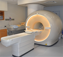
The Magnetic Resonance (MR) core is established to facilitate research and development within the Department of Radiology and collaborating departments in the field of MR imaging (MRI) and MR spectroscopy (MRS). The MR core consists of a whole-body human scanner (Philips Ingenia 3T MR) and a small animal Desktop scanner (Aspect 1T MR). Philips Ingenia includes dual-transmit and digital architecture for signal reception. This scanner enables the development and evaluation of new MRI/MRS techniques for improved diagnosis and understanding of the pathophysiology of disease. Aspect 1T scanner enables sequential imaging of small animals (e.g. mice and rats), without sacrificing them, for longitudinal monitoring of disease progression and/or therapy response. Being cited next to the small animal PET/CT and SPECT/CT scanners, this allows superposition of images for multimodality analysis.
Click here to explore more about the MR Core.
Small Animal Imaging Resource (SAIR)
- SAIR is an institutional facility that promotes and facilitates small animal imaging related to models of human disease with state-of-the-art equipment including depth resolved or planar optical imaging (fluorescence (FLI), bioluminescence (BLI) and chemiluminescence (CLI)), MRI, ultrasound, photoacoustic tomography, PET/CT, SPECT/CT and planar scintigraphy
- Infrastructure for animal handling (e.g., anesthesia, infusion, monitoring vital signs)
- Experienced investigators and technical staff capable of undertaking imaging and assisting in data interpretation are associated with the Resource and provide consultation on experimental planning, analysis and validation, and data archiving.
- Expertise in pulse programming and implementation for novel MRI experiments, design and acquisition or building of MR coils, choice of reporter molecules and /or genes, radiolabeling procedures and synthesis of ligands.
- Currently administered jointly by the Advanced Imaging Research Center (AIRC) and the Department of Radiology
Translational Molecular Imaging Core (TMIC)
- Cyclotron and radiochemistry facility approved for CGMP production of PET radiopharmaceuticals for human use. Capable of producing 6 radioisotopes and >30 radiotracers in addition to the FDA-approved tracers
- A regulatory office in the Department of Radiology facilitates Investigational New Drug (IND) and Abbreviated New Drug Applications (ANDA) approval of radiotracers.
- Radiochemistry and nuclear medicine experts to advise investigators on the development and implementation of imaging protocols in a range of disease models (e.g., cancer, diabetes, metabolism, cardiotoxicity, neurodegenerative diseases, etc.)
- Pre-clinical imaging including a Siemens Inveon PET/CT scanner for small animal imaging
- State-of-the-art human imaging in the NE2 building including a GE Discovery IQ five ring PET/CT scanner and a Siemens 3T Biograph hybrid PET/MR scanner, both located in close proximity to our cyclotron facility
- Publications