Spectral CT
Our Vision
Dual-Energy CT is a relatively new diagnostic imaging technology whose full clinical potential has yet to be achieved. By using customized software and Artificial Intelligence tools for image processing and analysis we have developed new methods that take full advantage of the material differentiation and quantification capabilities offered by Dual-Energy CT. These methods can be applied to both endogenous tissues and exogenous contrast agents. Overall, these methods allow us to discover new imaging biomarkers for disease detection and diagnosis with the potential to improve patient outcomes. The universality of these methods is shown by applying them to the Philips and Siemens scanners located at UT Southwestern and to GE scanners located at collaborating institutions.
Our Mission
To translate innovations in Dual-Energy CT imaging and technology to the clinic to optimize patient outcomes.
About Us
Our clinical research Program consists of a group of dedicated Radiologists and Scientists who have a common goal of maximizing the diagnostic information obtained from Dual-Energy CT scans. This new information allows us to find answers to biological questions that could not be answered with conventional CT scans.
- Faculty & Staff
Faculty & Staff

Assistant Professor

Assistant Professor
Medical Director, Computed Tomography
Assistant Professor

Assistant Professor

Professor
Vice Chair, Informatics
Assistant Professor

Assistant Professor
- Research Projects
Research Projects
Isoattenuating Gallstone Detection
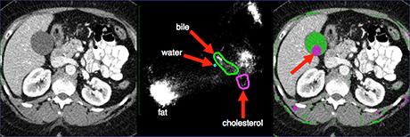
Approximately 15% of the adult population is affected by gallstones. Complications from gallstones include acute cholecystitis, biliary obstruction, pancreatitis, and even gallbladder carcinoma. Adding to this complication is the fact that 30% of all gallstones appear isoattenuating with bile in conventional CT images (see image above) making them difficult to detect. These patients often require further imaging with ultrasound or magnetic resonance (MRCP) that increases both the cost to the patient and the time to diagnosis. To meet this clinical need we have recently developed a Dual-Energy CT method that is capable of differentiating solid cholesterol gallstones from liquid bile (and other tissues) and segments them in a single image using colored pixels (see right image above). This allows the radiologist to quickly determine the exact location and size of gallstones that were previously invisible on conventional CT images, thus eliminating the need for subsequent imaging. For more information on this research project please contact Todd.Soesbe@UTSouthwestern.edu.
Liver Fat Quantification
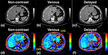
Non-alcoholic fatty liver disease (NAFLD) is highly prevalent in the United States where it affects one-third of the population. This silent epidemic usually remains asymptomatic until the late stages of the disease that include non-alcoholic steatohepatitis (NASH), fibrosis, cirrhosis, and even liver cancer (hepatocellular carcinoma). Currently, there are no approved therapies other than weight loss making early detection essential for positive patient outcomes. If the liver fat fraction could be included as a standard biometric in all abdominal CT scans regardless of iodinated contrast, then this would help to diagnose early-stage asymptomatic NAFLD. To meet this clinical need we have recently developed a Dual-Energy CT method capable of measuring the liver fat fraction in both non-contrast and contrast-enhanced scans. This method gives the same fat fraction regardless of the scan phase as seen in the conventional and fat fraction images above. For more information on this research project please contact Lakshmi Ananthakrishnan, M.D..
Artificial Intelligence
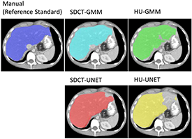
The additional x-ray attenuation information acquired by Dual-Energy CT is ideally suited for analysis using Artificial Intelligence where multiple layers of images can be used as the input. Convolutional Neural Networks like UNET that specialize in medical image processing can use this additional information to perform automatic liver segmentation (as shown above), removal of iodinated contrast (i.e., virtual non-contrast), isoattenuating gallstone detection, liver fat fraction measurements, and other opportunistic screenings.
Minimum-Iodine CTA
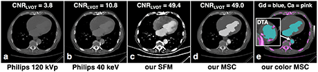
Contrast-enhanced CT examinations represent approximately half of all clinical CT scans and utilize the administration of intravenously injected iodinated contrast agents. Unfortunately, approximately 1% of all contrast-enhanced CT patients suffer from allergic-like or physiologic reactions due to these intravascular iodinated contrast agents. This represents a huge clinical problem for hundreds of thousands of patients who are in need of the diagnostic benefits of contrast-enhanced CT imaging but have risks associated with intravascular iodine. Therefore, creating safe options for these patients is a prevailing important clinical need. In order to meet this clinical need we have recently developed a Dual-Energy CT method that maximizes the conspicuity of intravascular contrast agents. With this method the true minimum dose of iodine contrast can be used, which is approximately one-tenth of a standard dose of iodine. Alternately, this method can also be used with gadolinium contrast using standard magnetic resonance doses (as shown above). For more information on this research project please contact Todd.Soesbe@UTSouthwestern.edu.
- Associated Cores
Associated Cores
DASPA
The Data Storage, Processing & Analysis core provides informatics support to researchers in a variety of areas.
Research PACS
- iPACS provides a HIPAA-compliant PACS system for archiving clinical and preclinical research imaging studies
- The installed and supported iPACS system is a web-based, secure, project-oriented resource available to Faculty researchers in Radiology and their collaborators
- Also has the capability of performing customized de-identification of images acquired in clinical trials to preserve patient confidentiality
ANSIR
The Advanced NeuroScience Imaging Research (ANSIR) lab dataset processing provides:
- Fully automated analysis of neuro MRI data including structural analysis using SPM and Freesurfer, task and resting state fMRI processing, Diffusion Tensor image processing, automated FLAIR white matter lesion segmentation, Arterial Spin Label MRI processing, and quantitative susceptibility mapping.
- Project-specific XNAT database storage allowing retrieval and queries of imaging and metadata
- Clinical alerts without a formal report can be provided for incidental findings
- Additional custom project-specific services may be available following consultation and based on resource availability
IM4T
The UTSW Radiology Department's Imaging Metrics for Trials (IM4T) group provides diagnostic imaging interpretation of cancer treatment response evaluation to internal and external researchers involved in cancer research studies that require response evaluation using Response Evaluation Criteria in Solid Tumors (RECIST) and its variants.
Magnetoencephalography (MEG)
- State-of-the-art technology mapping brain function
- Most advanced MEG technology currently available, and the only MEG scanner in Dallas
- Peripheral equipment available for time-locked stimuli and responses (ear buds, button pad, accelerometers, etc.)
- Used to study various neurological disorders and injuries including dementia, autism, concussion, and many others
- Publications
Publications
Soesbe TC, Xi Y, Nasr K, Leyendecker JR, Lenkinski RE, Lewis MA. Investigating new CT contrast agents: a phantom study exploring quantification and differentiation methods for high-Z elements using dual-energy CT. Eur Radiol. 2021 Apr 15. doi: 10.1007/s00330-021-07886-x. Epub ahead of print. PMID: 33856524.
Mileto A, Ananthakrishnan L, Morgan DE, Yeh BM, Marin D, Kambadakone AR. Clinical Implementation of Dual-Energy CT for Gastrointestinal Imaging. AJR Am J Roentgenol. 2020 Dec 30. doi: 10.2214/AJR.20.25093. Epub ahead of print. PMID: 33377415.
Duan X, Ananthakrishnan L, Guild JB, Xi Y, Rajiah P. Radiation doses and image quality of abdominal CT scans at different patient sizes using spectral detector CT scanner: a phantom and clinical study. Abdom Radiol (NY). 2020 Oct;45(10):3361-3368. doi: 10.1007/s00261-019-02247-1. PMID: 31587100.
Ng YS, Xi Y, Qian Y, Ananthakrishnan L, Soesbe TC, Lewis M, Lenkinski R, Fielding JR. Use of Spectral Detector Computed Tomography to Improve Liver Segmentation and Volumetry. J Comput Assist Tomogr. 2020 Mar/Apr;44(2):197-203. doi: 10.1097/RCT.0000000000000987. PMID: 32195798.
Soesbe TC, Lewis MA, Xi Y, Browning T, Ananthakrishnan L, Fielding JR, Lenkinski RE, Leyendecker JR. A Technique to Identify Isoattenuating Gallstones with Dual-Layer Spectral CT: An ex Vivo Phantom Study. Radiology. 2019 Aug;292(2):400-406. doi: 10.1148/radiol.2019190083. Epub 2019 Jul 2. PMID: 31264945.
Soesbe TC, Lewis MA, Nasr K, Ananthakrishnan L, Lenkinski RE. Separating High-Z Oral Contrast From Intravascular Iodine Contrast in an Animal Model Using Dual-Layer Spectral CT. Acad Radiol. 2019 Sep;26(9):1237-1244. doi: 10.1016/j.acra.2018.09.012. Epub 2018 Oct 9. PMID: 30314734.
Ananthakrishnan L, Duan X, Rajiah P, Soesbe TC, Lewis MA, Xi Y, Fielding JR, Lenkinski RE, Leyendecker JR, Abbara S. Phantom Validation of Spectral Detector Computed Tomography-Derived Virtual Monoenergetic, Virtual Noncontrast, and Iodine Quantification Images. J Comput Assist Tomogr. 2018 Nov/Dec;42(6):959-964. doi: 10.1097/RCT.0000000000000763. PMID: 29901508.
Ananthakrishnan L, Duan X, Xi Y, Lewis MA, Pearle MS, Antonelli JA, Goerne H, Kolitz EM, Abbara S, Lenkinski RE, Fielding JR, Leyendecker JR. Dual-layer spectral detector CT: non-inferiority assessment compared to dual-source dual- energy CT in discriminating uric acid from non-uric acid renal stones ex vivo. Abdom Radiol (NY). 2018 Nov;43(11):3075-3081. doi: 10.1007/s00261-018-1589-x. PMID: 29626256.
Megibow AJ, Kambadakone A, Ananthakrishnan L. Dual-Energy Computed Tomography: Image Acquisition, Processing, and Workflow. Radiol Clin North Am. 2018 Jul;56(4):507-520. doi: 10.1016/j.rcl.2018.03.001. PMID: 29936944.
Soesbe TC, Ananthakrishnan L, Lewis MA, Duan X, Nasr K, Xi Y, Abbara S, Leyendecker JR, Lenkinski RE. Pseudoenhancement effects on iodine quantification from dual-energy spectral CT systems: A multi-vendor phantom study regarding renal lesion characterization. Eur J Radiol. 2018 Aug;105:125-133. doi: 10.1016/j.ejrad.2018.06.002. Epub 2018 Jun 6. PMID: 30017268.
Kaza RK, Ananthakrishnan L, Kambadakone A, Platt JF. Update of Dual-Energy CT Applications in the Genitourinary Tract. AJR Am J Roentgenol. 2017 Jun;208(6):1185-1192. doi: 10.2214/AJR.16.17742. Epub 2017 Mar 16. PMID: 28301210.
Ananthakrishnan L, Rajiah P, Ahn R, Rassouli N, Xi Y, Soesbe TC, Lewis MA, Lenkinski RE, Leyendecker JR, Abbara S. Spectral detector CT-derived virtual non-contrast images: comparison of attenuation values with unenhanced CT. Abdom Radiol (NY). 2017 Mar;42(3):702-709. doi: 10.1007/s00261-016-1036-9. PMID: 28084546.