Image Reconstruction
Our Vision
The Reconstruction Program strives to leverage the full potential of state of the art hardware and acquisition methods to advance precision imaging, reduce scan time while maintaining image quality, and visualize new biomarkers both for human and preclinical subjects.
Our Mission
To improve both patient care and research through development and use of advanced reconstruction tools.
About Us
Advancing the program's mission requires collaboration of researchers with technical, clinical, and preclinical expertise. Our interdisciplinary team at UT Southwestern Medical Center partners with both outside academic institutions and industry to meet the program's objectives. Integral to these research efforts are the extensive in-house state of the art imagers and core facilities. The program's work spans the continuum from basic science developments, using simulations and phantom scans, to translational projects, exploring the potential of new reconstruction methods in a limited population, and clinical work involving hundreds of subjects that may directly benefit patient care.
- Faculty & Staff
Faculty & Staff
Assistant Professor
Research Lab
Assistant Professor
Assistant Professor

Associate Professor
Research Lab
Assistant Professor
- Research Projects
Research Projects
Positron Emission Tomography
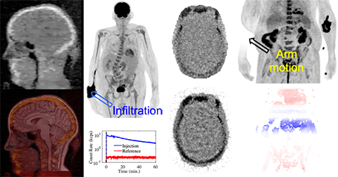
Generation of new reconstruction algorithms for PET kinetic modeling and improved data corrections (e.g. attenuation and motion) is being led by the Bowen Lab. The focus of these tools is to advance precision imaging for the care and study of oncology, neurology, and cardiology patients. The Bowen Lab also has expertise in vendor-provided offline reconstruction tools for both GE and Siemens PET scanners.
Magnetic Resonance Imaging
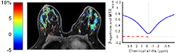
The Vinogradov Lab develops a range of post-processing tools to enable faster and more accurate chemical exchange saturation transfer (CEST) contrast imaging in-vivo. CEST is a novel contrast allowing visualization of important parameters that are inaccessible by current conventional MRI, such as pH or a particular metabolite level. The Madhuranthakam Lab leads the development of reconstruction algorithms for the following:
- Whole-body MRI for cancer screening and staging
- Fat/water separation
- Non-contrast perfusion imaging.
MultiSpectral Optoacoustic Tomography
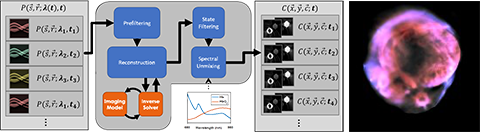
MultiSpectral Optoacoustic Tomography (MSOT) is an imaging modality which uses the differential absorption of near-infrared light to create images of the chemical constituents of a sample at high spatiotemporal resolution. The Mason Lab has produced the MATLAB Reconstruction, Analysis, and Filtering Toolbox (RAFT) for MSOT. The RAFT includes various methods for reconstruction and spectral unmixing, as well as offering multispectral state estimation to improve the temporal resolution and signal to noise ratio of analyzed datasets for improved dynamic imaging. It is parallelizable to improve processing throughput, and is available as a workflow on the BioHPC Astrocyte platform, enabling users without MATLAB to use the toolbox.
- Associated Cores
Associated Cores
DASPA
The Data Storage, Processing & Analysis core provides informatics support to researchers in a variety of areas.
Research PACS
- iPACS provides a HIPAA-compliant PACS system for archiving clinical and preclinical research imaging studies
- The installed and supported iPACS system is a web-based, secure, project-oriented resource available to Faculty researchers in Radiology and their collaborators
- Also has the capability of performing customized de-identification of images acquired in clinical trials to preserve patient confidentiality
ANSIR
The Advanced NeuroScience Imaging Research (ANSIR) lab dataset processing provides:
- Fully automated analysis of neuro MRI data including structural analysis using SPM and Freesurfer, task and resting state fMRI processing, Diffusion Tensor image processing, automated FLAIR white matter lesion segmentation, Arterial Spin Label MRI processing, and quantitative susceptibility mapping.
- Project-specific XNAT database storage allowing retrieval and queries of imaging and metadata
- Clinical alerts without a formal report can be provided for incidental findings
- Additional custom project-specific services may be available following consultation and based on resource availability
IM4T
The UTSW Radiology Department's Imaging Metrics for Trials (IM4T) group provides diagnostic imaging interpretation of cancer treatment response evaluation to internal and external researchers involved in cancer research studies that require response evaluation using Response Evaluation Criteria in Solid Tumors (RECIST) and its variants.
Magnetoencephalography (MEG)
- State-of-the-art technology mapping brain function
- Most advanced MEG technology currently available, and the only MEG scanner in Dallas
- Peripheral equipment available for time-locked stimuli and responses (ear buds, button pad, accelerometers, etc.)
- Used to study various neurological disorders and injuries including dementia, autism, concussion, and many others
Magnetic Resonance Imaging (MRI)
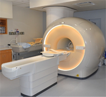
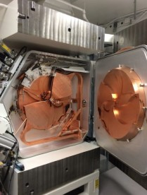
The Magnetic Resonance (MR) core is established to facilitate research and development within the Department of Radiology and collaborating departments in the field of MR imaging (MRI) and MR spectroscopy (MRS). The MR core consists of a whole-body human scanner (Philips Ingenia 3T MR) and a small animal Desktop scanner (Aspect 1T MR). Philips Ingenia includes dual-transmit and digital architecture for signal reception. This scanner enables the development and evaluation of new MRI/MRS techniques for improved diagnosis and understanding of the pathophysiology of disease. Aspect 1T scanner enables sequential imaging of small animals (e.g. mice and rats), without sacrificing them, for longitudinal monitoring of disease progression and/or therapy response. Being cited next to the small animal PET/CT and SPECT/CT scanners, this allows superposition of images for multimodality analysis.
Click here to explore more about the MR Core.
Small Animal Imaging Resource (SAIR)
- SAIR is an institutional facility that promotes and facilitates small animal imaging related to models of human disease with state-of-the-art equipment including depth resolved or planar optical imaging (fluorescence (FLI), bioluminescence (BLI) and chemiluminescence (CLI)), MRI, ultrasound, photoacoustic tomography, PET/CT, SPECT/CT and planar scintigraphy
- Infrastructure for animal handling (e.g., anesthesia, infusion, monitoring vital signs)
- Experienced investigators and technical staff capable of undertaking imaging and assisting in data interpretation are associated with the Resource and provide consultation on experimental planning, analysis and validation, and data archiving.
- Expertise in pulse programming and implementation for novel MRI experiments, design and acquisition or building of MR coils, choice of reporter molecules and /or genes, radiolabeling procedures and synthesis of ligands.
- Currently administered jointly by the Advanced Imaging Research Center (AIRC) and the Department of Radiology
Translational Molecular Imaging Core (TMIC)
- Cyclotron and radiochemistry facility approved for CGMP production of PET radiopharmaceuticals for human use. Capable of producing 6 radioisotopes and >30 radiotracers in addition to the FDA-approved tracers
- A regulatory office in the Department of Radiology facilitates Investigational New Drug (IND) and Abbreviated New Drug Applications (ANDA) approval of radiotracers.
- Radiochemistry and nuclear medicine experts to advise investigators on the development and implementation of imaging protocols in a range of disease models (e.g., cancer, diabetes, metabolism, cardiotoxicity, neurodegenerative diseases, etc.)
- Pre-clinical imaging including a Siemens Inveon PET/CT scanner for small animal imaging
- State-of-the-art human imaging in the NE2 building including a GE Discovery IQ five ring PET/CT scanner and a Siemens 3T Biograph hybrid PET/MR scanner, both located in close proximity to our cyclotron facility
- Associated Programs
Associated Programs
Advanced Imaging Research Center
The AIRC is a collaboration between UT Southwestern Medical Center and other North Texas institutions to further research in magnetic resonance imaging (MRI) and magnetic resonance spectroscopy (MRS), and translation of discoveries into clinical practice. The Center's research seeks to advance basic understanding, diagnostic techniques, and treatments for a wide range of diseases, including cancer, diabetes, obesity, Alzheimer's disease, schizophrenia, depression, autism, attention-deficit hyperactive disorder (ADHD), and diseases of the heart, lung, and liver.
Cyclotron & Radiochemistry
The Cyclotron and Radiochemistry Program is a UT Southwestern Medical Center-wide effort to develop the full capability of nuclear imaging, namely radioisotope-based imaging, for noninvasive assessment of physiological processes and abnormalities in animal models and in humans.
With a biomedical cyclotron and the capability to synthesize a variety of biomedical radioisotopes, this program leverages the cutting-edge technology of positron-emission tomography (PET) to enable discoveries that span multiple areas of medicine and physiology.
Visit the Cyclotron and Radiochemistry Program's website.
- Publications