
Planar Scintigraphy
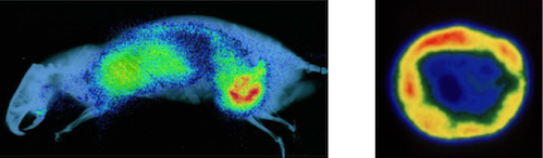
Whole-body planar scintigraphy of Dunning Prostate R3227-AT1 tumor-bearing rat. Left: Rat bearing 20 mm diameter tumor was injected IV with 5 MBq of [74As]bavituximab antibody. Anesthetized rat was imaged on a phosphor plate 72 h after injection and overlaid on a planar X-ray to provide anatomic correlation (Further details in Clin. Cancer Res., 14: 1377-1385 (2008) DOI: 10.1158/1078-0432.CCR-07-1516). Right: Autoradiography of slice of excised AT1 tumor following infusion of the vascular volume marker 125I-labeled albumin, revealing heterogeneity. Further details available in QJ Nucl. Med Mol Imaging 54(3):259-80 2010 [PMCID: PMC3044928; NIHMSID: NIHMS261584]