
PET/CT
These are some sample PET/CT images from the Small Animal Imaging Resource at UT Southwestern (UT-SAIR).
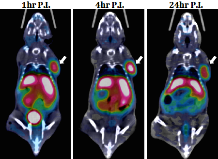
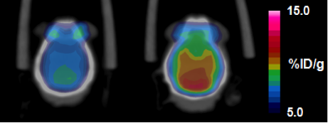
Brain tumor imaging: Representative coronal PET-CT images of 64Cu-c(DOTA-RGD), 64Cu-c(DOTA-RGDf), and 64Cu-DOTA-c(RGDyK) in U87MG tumor-bearing mice at 1 h, 4 h, and 24 h p.i. The white arrows indicate tumors. U87MG tumor model that has negative α vβ 3 and positive α 5β 1 integrins.
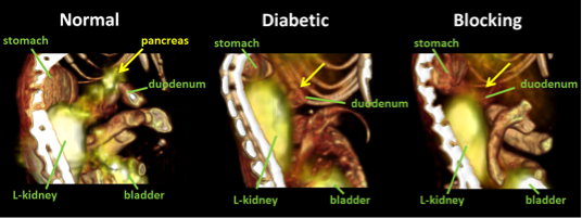
Imaging the pancreatic β-cell: These PET/CT images demonstrate that a 64Cu-labeled GLP-1 analog specifically target pancreatic beta cells in a mouse. Mice receiving the radiotracer showed a significantly elevated signal in the pancreas region as compared to mice co-injected with excess of cold EM2198 or in STZ-treated mice lacking beta cells. The imaging result was further confirmed by ex vivo imaging of the organs of interest excised from the mice and immunohistochemical staining of corresponding tissue slices.
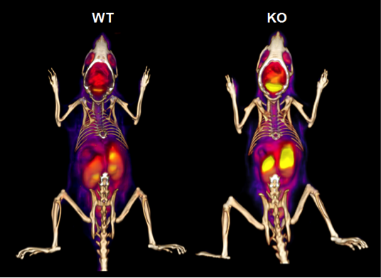
Imaging of brain glucose metabolism in ADHD: Quantitative dynamic PET images of FDG ( 18F-deoxyglucose) uptake into the brains of WT versus p35 KO mice demonstrate 50% greater uptake of glucose in the KO animals. This indicates that the absence of p35 results in an increase in glucose metabolism in brain. This study also demonstrates that small animal PET/CT is able to distinguish compartments within the brain and differences in the rate of glucose metabolism in those compartments may aid in diagnosis of hyperactivity disorders in patients.
Prostate cancer: FDG- PET-CT imaging of a mouse bearing PC-3-shDAB2IP tumors (7 days) at 1 h p.i. of 18F-FDG (100 μCi, i.v.). Ref: Xie et al. PNAS, 2010, 107(6): 2485-2490.