Instruments and Locations
The PCIRCF’s equipment is located as follows:
Inside the Barrier
NB3.206
- Optical imager
- MR
- SARRP irradiator
- PET
- CT
NG2.310
- X-Rad 320 Irradiator
Outside the Barrier
- NB3.406: X-Rad 320 Irradiator
- K1.406: X-Rad 320 Irradiator
- NF1.324: X-Rad 225Cx Irradiator
- NC7.122: XF24 Metabolic analyzer
Irradiation Devices
Small Animal Irradiation Platforms
The PCIRCF has two state-of-the-art small animal irradiators to deliver high-throughput, high-precision radiation experiments under image guidance:
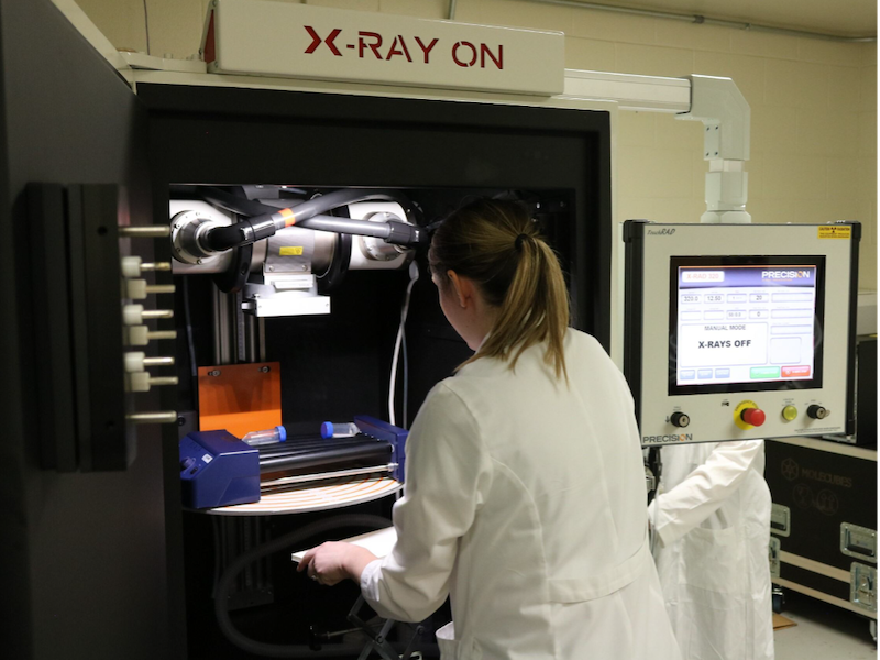
X-Rad 225Cx - NF1.324 (outside the animal facility barrier)
From Precision X-Ray Irradiation, Inc., the X-Rad 225Cx is a specialized radiation therapy system designed for small animal research. It provides high-precision radiation therapy with integrated image guidance and treatment planning capabilities, simulating clinical approaches for cancer treatment studies. Specifically, it enables accurate irradiation of lung and prostate models by combining conebeam CT imaging with targeted dose delivery. This system is instrumental for researchers studying radiation’s effects at the molecular level and assessing therapeutic responses in preclinical models.
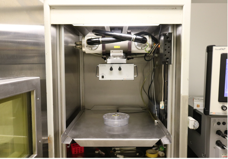
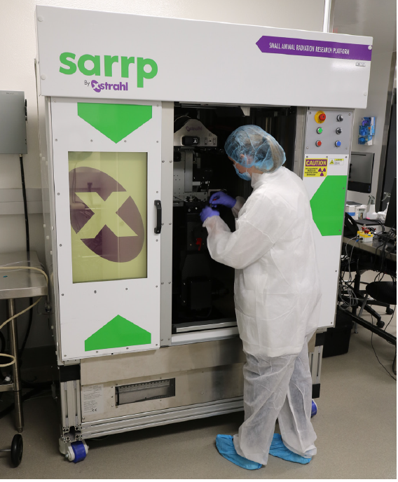
SAARP - NB3.206 (behind the animal facility barrier)
From Xstrahl Inc., the SAARP XStrahl irradiator is an advanced tool used in molecular radiation biology to deliver controlled X-ray radiation to biological specimens. It offers several innovative features that enhance research capabilities:
- 4D gating: Allows researchers to synchronize radiation delivery with the motion of the specimen, such as breathing in small animals. This precision minimizes damage to healthy tissues and improves the accuracy of radiation treatment, making it ideal for studying dynamic biological processes.
- Cone beam computed tomography (CBCT) imaging: Provides high-resolution, 3D images of the specimen before, during, and after irradiation. This capability enables researchers to visualize anatomical structures and assess radiation effects on tissues, facilitating more informed experimental planning and analysis.
- MicroCT imaging: Allows the irradiator to produce detailed, high-resolution images of small biological samples. It is particularly useful for examining the internal structures of small organisms and tissues at a microscopic level, allowing for in-depth analysis of radiation-induced changes.
X-Ray Irradiators
The PCIRCF has recently acquired two high-dose-rate X-ray irradiators for delivering radiation to small animals and biological specimens. These irradiators are primarily used for total body irradiation of mice, as well as radiation exposure for flies and cells. The PCIRCF will offer training, dosimetry services, and ongoing equipment maintenance.
X-RAD 320 - NB3.406 and K1.406 (outside the animal facility barrier) and NG2.310 (inside the barrier)
From Precision X-Ray Irradiation Inc., the XRad320 X-ray irradiator is a versatile device commonly used for delivering controlled doses of x-ray radiation to biological specimens. Its adjustable dose rates and precision targeting make it ideal for accurate dosing in animal and cell studies. The XRad320 is particularly suited for:
- Mouse and rat total body and focal radiation: Can perform whole-body irradiation of mice, making it valuable for studying radiation’s effects on the entire organism, modeling radiation exposure scenarios, and researching cancer therapies.
- Fly radiation: Used to expose flies to X-rays, allowing researchers to study genetic mutations, developmental changes, and survival rates in response to radiation, often for genetic and aging studies.
- Cell radiation: Widely used to irradiate cell cultures, facilitating studies on DNA damage, repair mechanisms, cell death, and cellular responses to ionizing radiation.
Imaging Devices
The PCIRCF has the following imaging devices to meet different needs:
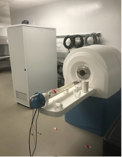
MRS 3017 - NB3.206 (behind the animal facility barrier)
The MRS 3017 3T MRI System, an MRI scanner with a 3T magnet field and 17 cm bore size, enables high-resolution imaging of target areas and fiducials -- small markers used for precise localization. This capability is directly applicable to generating detailed phantom images for pre-treatment validation, ensuring precision in therapy planning.
Overall, close proximity between an MRI and an irradiator provides significant advantages:
- Streamlined workflows
- Facilitated accurate tumor localization
- More effective and personalized patient treatments
With an integrated setup, the treatment planning system can seamlessly load CBCT data, overlay it with MRI data, and conduct image registration to align fiducials accurately. This improves target identification and planning, as the MRI’s soft tissue contrast enhances the precision of the treatment area.
SAARP - NB3.206
The SAARP irradiator allows for 4D gating and high-resolution imaging for precise radiation studies on small specimens. Additionally, the close proximity of the SAARP XStrahl irradiator and the MRS 3T MRI system enhances treatment precision by facilitating seamless integration of imaging and radiation delivery.
- System is equipped with both cone-beam CT and micro-CT systems for imaged-guided radiation therapy, as well as respiratory gating assistance for precise radiation therapy
X-RAD 225Cx - NF1.326
- System is equipped with a cone-beam CT for image-guided radiation therapy
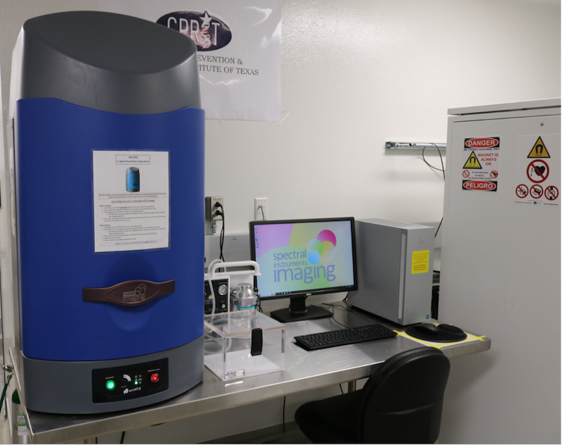
Ami HTX Optical Imaging System (NB3.206)
The Ami HTX Optical Imaging System is designed for non-invasive in vivo imaging of small animals, primarily mice. This system supports various imaging modalities that enable the monitoring of biological processes and disease progression at the molecular level. It also aids in visualizing how cells respond to radiation at the molecular level, tracking DNA repair mechanisms, and assessing the effectiveness of radiation therapies by monitoring tumor response in real-time. Key features of this imaging system include:
- Bioluminescent imaging
- Fluorescent imaging
- Near-infrared imaging
- X-ray imaging
- Five-mice platform
Alongside a small animal PET/CT, this dual system enables the assessment of tumor growth, tracking radiolabeled biomarkers for DNA repair and hypoxia, and understanding tissue responses to radiation, aiding in personalized therapy development, and exploring radiation resistance.
System specifications include:
- Bioluminescent imaging
- Fluorescent imaging
- X-ray imaging
- High throughput
- Pure LED illumination
- 100X light intensity on specimen
- 10 LED wavelengths from 430nm to 745nm
- 10 filters included from 530nm to 790 nm
- Solid state cooled CCD camera (–90C)
- High-performance imager
- CCD camera with back illumination
- Ultra-wide category leading 25cm x 17cm optical FOV
- X-ray FOV is 25 x 15 cm
- Up to 40kV maximum X-ray energy
Molecubes - small animal PET/CT NB3.206
Molecubes are compact, high-performance imaging systems designed for preclinical research. They are modular devices that combine multiple imaging modalities, such as PET (Positron Emission Tomography), SPECT (Single Photon Emission Computed Tomography), and CT (Computed Tomography), into one system, allowing researchers to obtain detailed images of small animals like mice and rats.
Metabolic Analyzer
Seahorse XFe24 Analyzer - NC7.122
The XFe24 Seahorse Extracellular Flux Analyzer measures cellular bioenergetics, focusing on oxygen consumption rate and extracellular acidification rate. These measurements give insights into cellular metabolic activities, such as mitochondrial respiration and glycolysis.
When cells are exposed to radiation, they undergo metabolic changes that impact their survival, proliferation, and response to therapy. By using the XFe24 analyzer, researchers can observe shifts in metabolic pathways and identify biomarkers or metabolic vulnerabilities. These insights facilitate development of new therapeutic strategies, especially for radiotherapy, by targeting metabolic pathways to improve the effectiveness of treatments while minimizing damage to healthy tissue.
The Seahorse Prep Station standardizes cell preparation for Seahorse Extracellular Flux Analyzers, automating steps like cell plating and media changes. This ensures consistent and precise measurements of metabolic parameters, which are critical for studies on cellular responses to treatments such as radiation in molecular biology and oncology.
Computational Instrumentation
Our facility includes a computational center that offers precise dosimetry and treatment planning, utilizing advanced Monte Carlo simulation tools on a high-performance GPU platform.
Quality Assurance
The PCIRCF maintains a quality assurance program, with physics personnel conducting weekly, quarterly, and annual assessments on all equipment to validate and maintain both geometric and dosimetric accuracy. Dosimetric measurements, performed using devices with accuracy traceable to national standards, ensure that the dosimetric uncertainty of our irradiators remains within ±3%.