MPE Facilities and Equipment
Hardware
Small Animal Irradiator
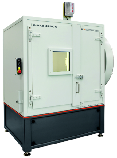
Animal irradiation systems facilitate scientific testing of biomedical hypotheses in vitro (cell) and in vivo (live animals) with the ultimate aim of promoting translational research and provide novel protocols for human cancer treatments. UT Southwestern uses an X-RAD 225Cx (Precision X-Ray Inc., N. Branford, CT) irradiator as the small-animal platform. The XRAD 225Cx irradiator consists of four main components: an industrial X-ray unit, a small field collimating system, a c-arm rotating gantry and an image guidance system. All components are enclosed in a self-shielding cabinet providing operation without the need for special shielding. The irradiator employs a dual focal spot X-ray tube, capable of both on-board imaging (30-100 kVp) for image-guided targeting and high precision treatment delivery (225 kVp). A robust gantry system, capable of a full 360-degree rotation, provides consistent geometry thanks to a reinforcing gantry ring. A motorized stage with three axes of translation can place an animal with submillimeter positional accuracy. The system’s collimators can modulate the irradiation from large 40x40 mm square fields down to 1 mm diameter circular beams. This allows for a large variation in possible dose patterns, including irradiating multiple targets within one specimen
3D Printer
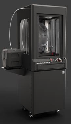
This commercial 3D printer is able to print extra-large (30.0 L x 30.5 W x 45.7 H cm) prototypes, models, and products. The printer uses Fused Deposition Modelling (FDM) technology. Polylactic acid (PLA) filament is fed into and melted by a heated extruder nozzle. The melted plastic then extrudes out and deposits at predetermined locations layer by layer. The plastic quickly hardens and forms into the designed shape. This printer is currently used to produce patient-specific boluses for radiation therapy. We have developed a clinical workflow from patient CT simulation to bolus manufacturing. Other examples of the prints that we produced are: custom brachytherapy QA phantom, breast phantoms, breast immobilization cups, etc.
GPUs
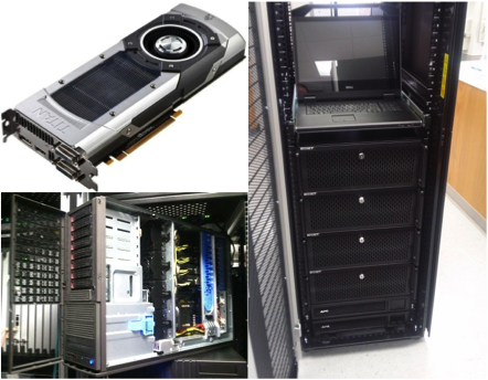
Graphics processing units are a relatively new type of high-performance computing device. Originally developed to handle computer graphics-related problems, GPUs are increasingly employed in high-performance scientific computing due to their tremendous parallel processing power. At UTSW, we have been developing GPU-based computational tools for radiotherapy. To support these activities, GPUs are installed on almost every research computer workstation for research development. A GPU cluster is also available in our department to enable large-scale computations needed to tackle challenging tasks. The cluster currently contains 37 GPU cards from major vendors, e.g. Nvidia Titan Black card. The cluster is housed in the department computer server room and maintained by our IT group. Research members remotely access those workstations via high-speed Internet. In addition, the GPU cluster also contains several Intel Zion Phi processors.
Research Workstations
Treatment planning systems (TPS), the "Grand Central" of imaging and therapy information transfer, is the heart of the radiation therapy process. Due to the rapid development of new planning technologies on the research side, we utilize dedicated research TPS to bridge the gap between new ideas and clinical implementations. The research TPS features the most updated commercialized planning technologies (knowledge-based planning technology, 4D planning, Monte Carlo dose calculation algorithms, VMAT, biological optimization, etc.) and advanced scripting capabilities allowing developers to write scripts to access the treatment planning information. The scripts and the resulting plans can be integrated into the clinical delivery systems for real delivery tests and dose evaluations. We currently have four research TPS serving both clinically innovative dosimetry projects and development of novel radiation therapy technologies.
We also use Velocity as a research tool. Velocity is a 3D imaging software program providing the means to display, register, and segment medical image volumes from multi-modality sources including CT, MRI, PET, and SPECT images. It features multi-modality image registration, automated contouring, and advanced treatment plan reviewing. Velocity image registration platform allows users to integrate all images and treatment plans and combine dose delivered by all treatment modalities. The contour platform provides an intuitive interface to ease tedious contouring tasks. The automated segmentation algorithms can create anatomical structures in seconds. The treatment planning evaluation platform simplifies modern radiation therapy plan review and evaluation across treatment platforms such as cyberknife, tomotherapy and brachytherapy. Treatment plan dose can be viewed using isodose curves and dose volume histograms. While being routinely used in clinic, Velocity also serves as a useful research platform, helping researchers on evaluating and validating self-developed algorithms.
Research Phantoms
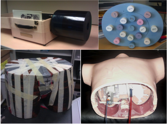
A wide variety of physical phantoms are available in our division to facilitate various experimental studies. These include Catphan phantom and Rando anthropomorphic phantom for CT imaging studies, electron density phantom for linac calibrations, CIRS lung phantom for imaging and dosimetric studies, and a motion platform for 4D motion related projects, etc.
Software
DICOManTX
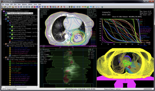
DICOManTX is a software system developed for users in medicine who need to handle DICOM objects especially DICOM RT (Radiation Therapy) objects. There are many DICOM applications that users can download and use free of charge, but they are mostly limited to image objects. It is very hard, if not impossible, to find a free application that is capable of managing and maneuvering DICOM RT objects, i.e., RT Image, RT Structure Set, RT Plan, RT Dose and RT Record objects, in a way that users in Radiation Therapy are used to. First devised in 1997, the five aforementioned RT-specific classes of objects are extensions to DICOM standards to model treatment workflows in clinics of Radiation Therapy. Most of them are non-image objects that are intertwined and connected in either internal or external links. RT expertise is needed to comprehend those objects. It is even more so for application developers. DICOManTX was designed in such a way that it would benefit a wide spectrum of users from newbies to the advanced. It is an all-in-one DICOM wizard which provides the user with DICOM RT viewer, Pusher, Retriever, Editor, Storage Server, Format Converter and so on. It presents DICOM objects in a structure that was defined by the RT information standard. So it is a useful gadget for education and R&D activities. More RT-specific functions are being constantly added. It is also becoming a peer-review tool for clinical trials in Radiation Therapy.
SCORE
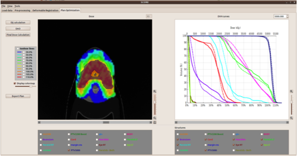
An online photon ART platform, SuperComputing for Online Replanning Environment (SCORE), has been developed at UTSW. It includes a user-friendly graphical user interface (GUI), a set of GPU-based real-time re-planning tools, and an interface with commercial treatment planning systems (TPS). The GUI is developed using Qt, a cross-platform application development framework. Under this GUI, we can load the initial treatment plan in DICOM-RT format from a commercial TPS and a CBCT image from an imaging console. To perform treatment replanning, a number of preprocessing procedures on the CBCT image are applied first, such as rigid registration with the planning CT and removal of CT couch. Deformable image registration is then performed using demon's algorithm to transfer the contours from the planning CT to the CBCT. These contours on the CBCT will be inspected and, if needed, manually modified by the user. After that, the treatment plan will be re-optimized and final dose distribution and DVH can be calculated and displayed. Finally, the re-optimized plan is output in DICOM-RT format and transferred to the commercial TPS. For computationally intensive steps, SCORE invokes corresponding GPU modules to achieve high efficiency. The SCORE system serves as the platform for a number of treatment optimization projects, e.g. automatic treatment planning and interactive treatment planning.
GPU-MC Toolkit
The Monte Carlo (MC) technique is commonly considered as the most accurate method for radiation transport simulation in radiotherapy. It has been widely used in many problems ranging from radiation dose calculation to CT imaging simulation. However, its efficiency hinders their research and clinical applications. We have developed a set of GPU-based MC particle transport simulation packages for different particle types, energy ranges, and clinical applications:
- gDPM and goMC: mega-electron volt energy range coupled photon-electron transport simulations for external beam radiotherapy dose calculations.
- gCTD and gMCDRR: kilo-electron volt energy range photon transport simulation for x-ray based medical imaging simulations and dose calculations.
- gBMC: photon transport simulation for brachy therapy dose calculation.
- gPMC: transport simulation of proton and secondary particles for proton therapy dose and linear energy transfer calculation.
- gCMC: transport simulation of carbon ion and secondary particles for carbon therapy applications.
These GPU-enabled MC packages utilize parallel computation ability of a GPU to achieve high efficiency (typically ~100 times accelerations over single CPU-based MC simulations), while employing appropriate particle transport physics and hence maintaining simulation accuracy. We have developed various techniques to achieve efficient parallel processing under GPU’s single instruction multiple data processing scheme. A few of the recently developed packages were based on OpenCL platform, which substantially improved code's portability, allowing them to be executed on different platforms including conventional CPUs and GPUs from different vendors.
GPU-Radiotherapy Toolkit
Over years, a number of research codes and software packages in radiotherapy have been developed, forming a GPU-radiotherapy toolkit. The development of this toolkit aims at solving computationally intensive tasks with high computational efficiency and user-friendly interface. These codes are now widely used by all the department members. The use of these high quality packages greatly simplifies and speeds up the research development process. Modules inside the toolkit include:
- Pencil beam dose calculation: Finite-size pencil beam dose calculation tool for photon therapy with 3D density correction to achieve accuracy.
- Inverse treatment plan optimization: A set of inverse optimization tools to solve fluence map optimization, direct aperture optimization, and volumetric arc therapy optimization.
- γ-index dose comparison: ultra-fast 2D and 3D γ-index calculations using a geometric search method with a radial pre-sorting technique.
GPU-Image Processing Toolkit
Medical imaging is a core component of radiotherapy. This GPU-based imaging processing toolkit contains a number of frequently used modules in typical imaging problems, such as 2D/3D image denoising, deformable image registration, and CT reconstruction.
- Image denoising: advanced image denoising methods solved with iterative algorithms, such as total variation, tight frame, nonlocal means, etc.
- Image Registration: both rigid and non-rigid (deformable) image registration. The deformable registration tool gDEMONS implements demons DIR algorithm with a multi-scale scheme on GPU.
- Forward and backward (cone beam) CT projection: forward and backward projection calculations in a (cone beam) CT geometry realized via Siddons algorithm, trilinear interpolation algorithm, etc.