
Hyperion Imaging System - Spatial Analysis
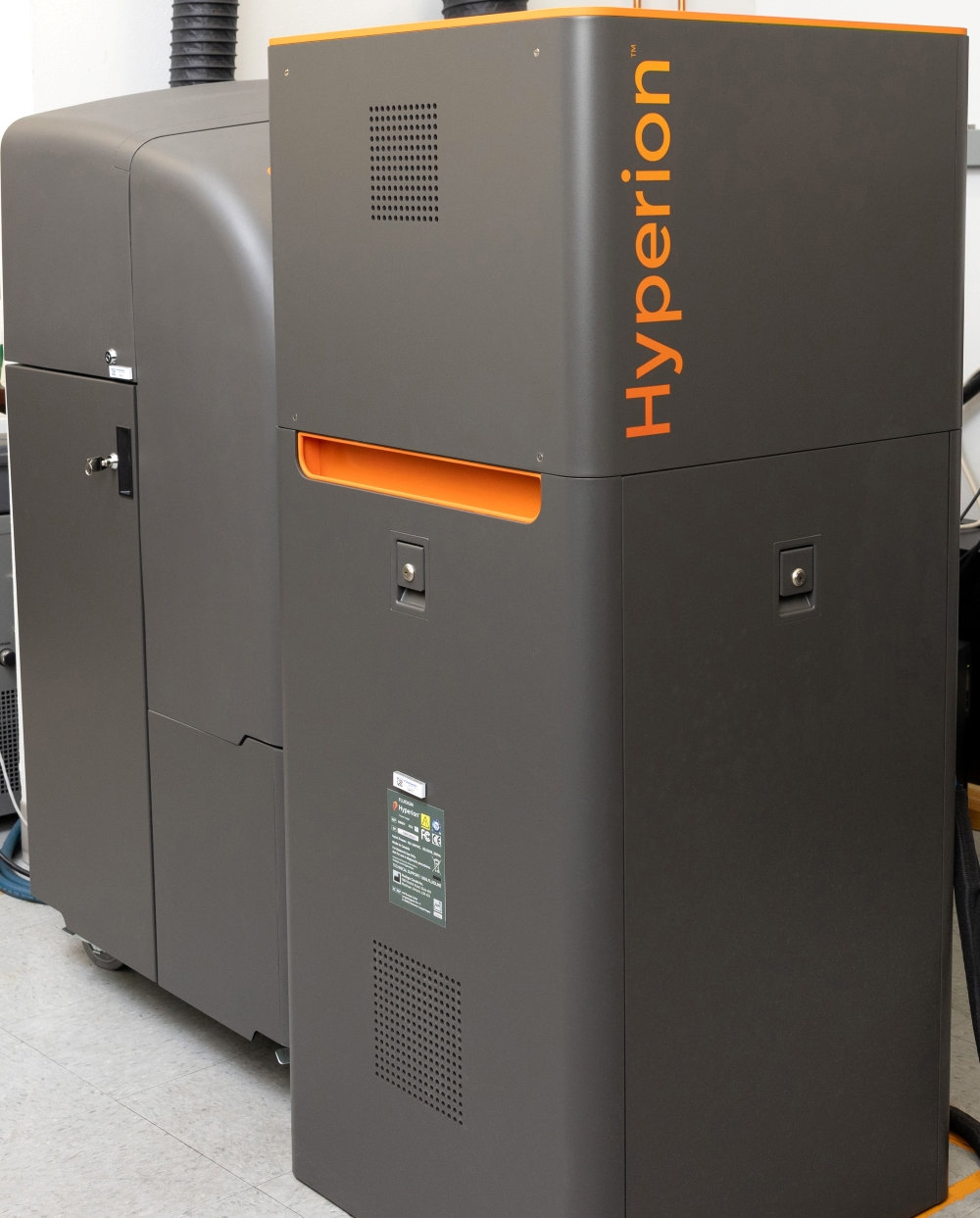
The Hyperion Imaging System extends mass cytometry to tissue analysis by combining metal-tag detection with laser ablation.
Best suited for:
- Spatial profiling of tissue sections
- Tumor microenvironment analysis
- Cell–cell interaction and tissue architecture studies
This platform enables simultaneous detection of 40+ markers while preserving spatial context, allowing researchers to understand not only what cells are present, but where they are located and how they interact.
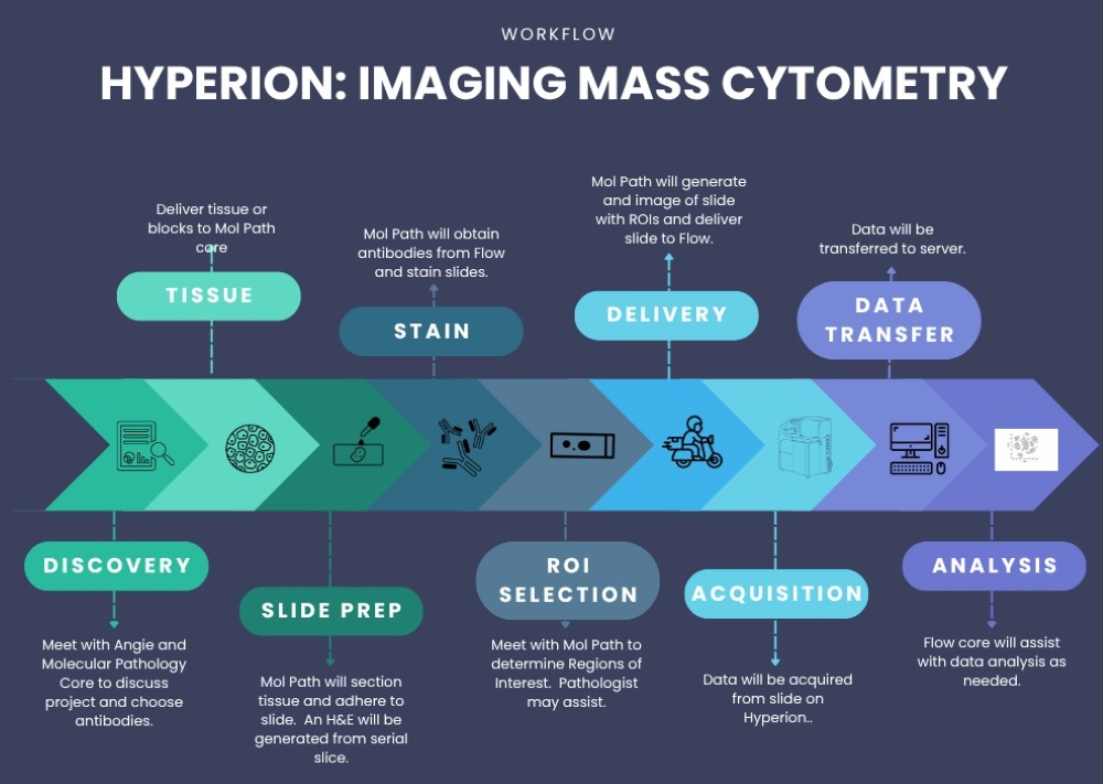
Mass Cytometry Workflow Process
Need help getting started or planning your experiment?
Angela.mobley@utsouthwestern.edu Angela Mobley for expert guidance.