UT Southwestern stem cell biologists develop embryo model
Peri-gastruloids advance understanding of early human development, tissue formation, and differentiation for regenerative medicine
DALLAS – July 20, 2023 – UT Southwestern Medical Center biologists have developed a new stem cell-based embryo model for studying early human development, tissue formation, and differentiation, offering valuable contributions to the field of developmental biology and regenerative medicine.
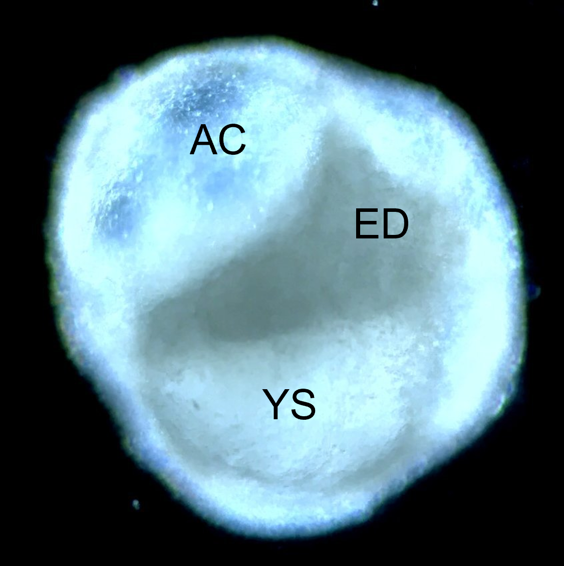
Peri-gastruloids are primitive embryonic structures with the potential to form many of the different cell types of the body, thereby providing a window into the earliest steps of embryogenesis. The findings on peri-gastruloids, published in Cell, could impact a wide range of diseases and conditions that involve early human development, including developmental disorders, birth defects, genetic disorders, and diseases that arise during embryonic development such as certain types of cancer or neurological disorders, researchers noted. Disease modeling and drug testing also can benefit from peri-gastruloid research.
“By generating peri-gastruloids, researchers can mimic and study the complex processes that occur during gastrulation and early organ formation, which is a critical period in embryonic development. This allows for a better understanding of the molecular and cellular events that shape the formation of different tissue layers,” explained Jun Wu, Ph.D., Assistant Professor of Molecular Biology, Virginia Murchison Linthicum Scholar in Medical Research, and member of the Harold C. Simmons Comprehensive Cancer Center at UT Southwestern.
“Studying peri-gastruloids allows researchers to gain insights into how abnormalities or genetic mutations can impact the early stages of human development. This knowledge can be used to model and understand developmental disorders or diseases that arise during early embryonic development. Additionally, peri-gastruloids can serve as a platform for testing the efficacy and safety of potential drugs or therapies targeting these conditions,” Dr. Wu said.
“The ability to model human development using peri-gastruloids can provide insights into the mechanisms behind tissue formation and differentiation. This knowledge can be applied to improve regenerative medicine approaches, such as generating specific cell types or tissues for transplantation or repairing damaged organs,” said lead author Lizhong Liu, Ph.D., Assistant Instructor of Molecular Biology in the Wu lab, which focuses on using stem cell models to learn more about mammalian development and create regenerative medical applications.
While in vitro stem cell models that recapitulate human gastrulation have been established, they lack the essential extraembryonic cells needed for embryonic development, morphogenesis, and patterning. The new approach provides a robust and efficient method that prompts human pluripotent stem cells to self-organize into peri-gastruloids, which encompass both embryonic (epiblast) and extraembryonic (hypoblast) tissues. Although peri-gastruloids are not viable due to the exclusion of trophoblasts, they can simulate critical stages of human peri-gastrulation development, such as forming amniotic and yolk sac cavities, developing bilaminar and trilaminar embryonic discs, specifying primordial germ cells, initiating formation of primary germ layers, and early organogenesis.
By replicating conditions in vitro, researchers can delve into the underlying mechanisms and identify potential therapeutic targets for the eventual development of novel treatments or interventions for patients. Researchers also can expose peri-gastruloids to various drugs or compounds to evaluate their efficacy and potential side effects. This can help in identifying promising drug candidates and ensuring the safety of pharmaceutical interventions before they are tested in human clinical trials.
“Overall, the study’s findings on peri-gastruloids contribute to the advancement of clinical solutions by providing a platform for regenerative medicine, disease modeling, and drug testing, which have the potential to lead to the development of new therapies, improved treatment strategies, and enhanced safety assessment methods for the benefit of patients,” said Dr. Wu, who is a Cancer Prevention and Research Institute of Texas (CPRIT) Scholar and a New York Stem Cell Foundation (NYSCF) – Robertson Investigator.
Generating new stem cells and stem cell-based embryo models to study embryogenesis and developing translational applications using stem cells represent the central directions of the Wu lab. The lab currently focuses on deriving novel pluripotent stem cells, identifying and overcoming the xenogeneic barriers, studying novel regulators of pluripotency, and generating stem cell-based models of mammalian embryos.
Among his published research, Dr. Wu has:
- Generated biological structures that resemble human blastocysts, the structures that arise from the early development of fertilized eggs in mammals, and used them to investigate human pluripotent stem cells, which have the potential to become nearly all of the body’s many tissue types.
- Developed a method to produce bovine blastoids, a crucial step in replicating embryo formation in the lab that could lead to the development of new reproductive technologies for cattle breeding.
As part of the Wu Lab, Dr. Liu's research utilizes pluripotent stem cell-based 3D embryo models to study peri-gastrulation, aiming to expand understanding of early development particularly encompassing the specification of primary germ layers, neurulation, and organogenesis. His ultimate goal is to build knowledge that might mitigate early pregnancy loss and congenital defects, and help to generate lab-grown human tissues and organs for applications in regenerative medicine.
The research was conducted with collaboration from the Hamon Center for Regenerative Science and Medicine and Cecil H. and Ida Green Center for Reproductive Biology Sciences at UT Southwestern, and the Institute for Regenerative Medicine at the Perelman School of Medicine, University of Pennsylvania, along with researchers from the Department of Obstetrics and Gynecology and the Lyda Hill Department of Bioinformatics at UT Southwestern.
Other UTSW researchers who contributed to this study include Seiya Oura, Ph.D., postdoctoral research fellow; graduate students Zachary Markham, James Hamilton, and Carlos A. Pinzon Arteaga; Leijie Li, incoming postdoctoral research fellow; Masahiro Sakurai, Ph.D., Assistant Instructor; Lei Wang, Ph.D., Research Scientist; and Gary C. Hon, Ph.D., Associate Professor.
About UT Southwestern Medical Center
UT Southwestern, one of the nation’s premier academic medical centers, integrates pioneering biomedical research with exceptional clinical care and education. The institution’s faculty members have received six Nobel Prizes and include 24 members of the National Academy of Sciences, 25 members of the National Academy of Medicine, and 13 Howard Hughes Medical Institute Investigators. The full-time faculty of more than 3,300 is responsible for groundbreaking medical advances and is committed to translating science-driven research quickly to new clinical treatments. UT Southwestern physicians in more than 80 specialties care for more than 143,000 hospitalized patients, attend to more than 470,000 emergency room cases, and oversee nearly 5.3 million outpatient visits a year.