Study links female sex hormones to progression of eye disease
UTSW findings could lead to treatments to slow vision loss from retinitis pigmentosa
DALLAS – July 08, 2025 – Female sex hormones can significantly enhance the progression of the rare neurodegenerative eye disease retinitis pigmentosa (RP), according to a preclinical study by researchers at UT Southwestern Medical Center. The findings, published in Science Advances, may lead to therapeutics to slow progression of the disease and help clinicians assess the risk of hormone therapies for female patients with genetic markers for the disease.

“Retinitis pigmentosa is a commonly inherited form of vision loss that was previously thought to be unaffected by biological sex,” explained Katherine Wert, Ph.D., Assistant Professor of Ophthalmology at UT Southwestern. “We discovered we can actually slow disease progression in female mice if we deplete their sex hormones.”
The eye disease, which can lead to blindness, affects about 1 in 3,500 people in the U.S. and causes photoreceptors – light-sensitive cells in the retina – to degenerate and die. It is linked to over 75 genes and can occur in people of any age. There is currently no cure.
Dr. Wert noted that previous research indicated a connection between sex and other degenerative retinal conditions, but this is the first study to establish hormone signaling as a direct cause.
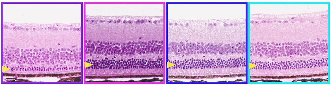
The researchers studied male and female mice with a mutation in rhodopsin (RHO P23H) – a key light-sensing protein in the retina – that causes the development of retinitis pigmentosa. At 2 months of age, they found the layer of photoreceptors in the females’ retinas had significantly worse function than in males, indicating faster vision loss and a sex-related difference in disease progression.
In females spayed to reduce sex hormone levels, Dr. Wert said the team was surprised to see reduced RP progression, to the point that their vision was comparable to that of males. Neutered males showed no change in visual function compared to intact males. Similarly, artificially increasing sex hormone levels in neutered mice by implanting estradiol under their skin had no effect on males but resulted in increased loss of function in the photoreceptors of females.
In contrast, the vision of healthy mice without the RHO P23H mutation remained unchanged after they received female sex hormones, indicating an interaction between the rhodopsin mutation and sex hormones, causing higher levels of inflammation and cell death in females.

Ashley Rowe, the study’s lead author and a doctoral student in Dr. Wert’s laboratory, noted that the team’s research warrants further investigation regarding the potential impact of hormonal therapy in women with related retinal conditions. The authors emphasized that there is currently no evidence that birth control and hormone replacement therapy adversely affect vision in women without genetic mutations associated with retinitis pigmentosa.
“We’re not advocating for depleting women’s hormones,” Ms. Rowe said. “But this discovery opens exciting opportunities to understand the underlying mechanisms leading to worsened disease outcomes in women, which could lead to transformative future therapeutic strategies for both women and men living with RP.”
Other UTSW researchers who contributed to the study are Jeffrey McDonald, Ph.D., Professor of Molecular Genetics; Mauricio Velasquez, B.S., a Senior Analytical Chemist in the Center for Human Nutrition and the McDonald Lab; and Wert Lab members Jacob Aumeier, medical student, and Tiffany Yee and Emily Nettesheim, Ophthalmology doctoral students.
This research was funded by grants from the National Institutes of Health (R21EY034597, P30EY030413, 5T32GM131945, P01HL-160487, P30DK127984, UL1TR003163, F31EY036730), a Research to Prevent Blindness Challenge Grant, and the Van Sickle Family Foundation.
About UT Southwestern Medical Center
UT Southwestern, one of the nation’s premier academic medical centers, integrates pioneering biomedical research with exceptional clinical care and education. The institution’s faculty members have received six Nobel Prizes and include 24 members of the National Academy of Sciences, 25 members of the National Academy of Medicine, and 13 Howard Hughes Medical Institute Investigators. The full-time faculty of more than 3,300 is responsible for groundbreaking medical advances and is committed to translating science-driven research quickly to new clinical treatments. UT Southwestern physicians in more than 80 specialties care for more than 143,000 hospitalized patients, attend to more than 470,000 emergency room cases, and oversee nearly 5.3 million outpatient visits a year.