News
Click on the news sections to expand:
- 05/2026: New Leica Stellaris Falcon 8
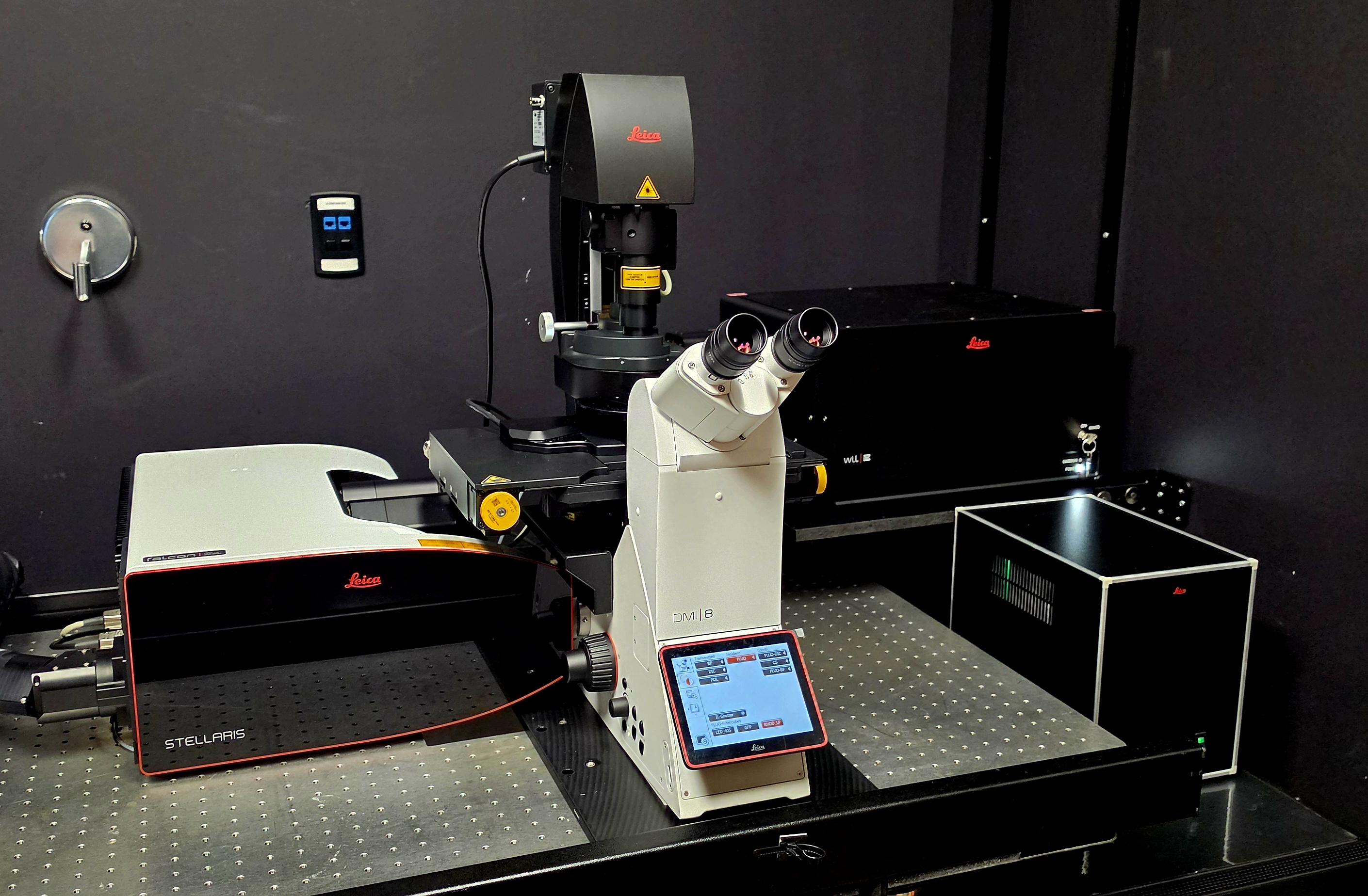
Leica Stellaris Falcon 8
The QLMC is excited to introduce the Leica Stellaris Falcon 8 to the UTSW research community! The Leica Stellaris Falcon 8 is a confocal point scanning system with White Light Laser (WLL) as excitation light source and a highly sensitive, prism-based spectral detection design with computer controlled adjustable bandwidth for all fluorescence channels. The extended detection range up to 850 nm, plus the expanded excitation range in the visible from 440 nm up to 790 nm allows for the excitation and detection of fluorescent probes with the highest specificity. Combined with TauSense, the system offers a set of tools based on fluorescence lifetime information, which provide additional contrast, improve image quality and separation of spectrally overlapping fluorophores. The system features a tandem scanning head to provide highest axial resolution with galvanometric scanners that are switchable between resonant and non-resonant mode. The Leica STELLARIS FALCON system provides fast FLIM (at confocal speed) with any imaging workflow. Quick and easy operation is ensured by the full integration within LAS X. STELLARIS FALCON includes FLIM analysis tools, such as exponential fitting, FLIM phasor analysis, pattern fitting and FLIM-FRET analyzer.
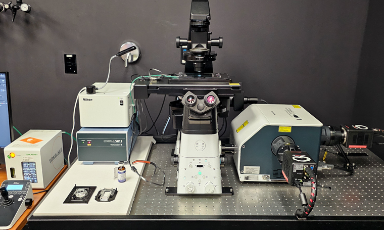
- 04/2026: Nikon CSU-W1 dual cam gets a new home
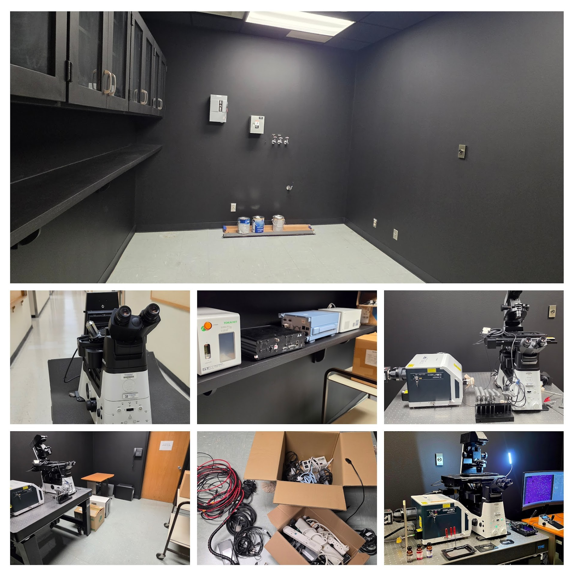
Nikon CSU-W1 dual cam moves to South campus
The QLMC currently only operates one Zeiss LSM880 with AiryScan on South Campus. As a result, all users requiring high temporal resolution need to transport their samples to North campus to take advantage of our spinning disk confocals. Furthermore, on North campus, all available rooms/slots are filled with microscopes. This means there is currently no space for the installation of our new Leica Stellaris Falcon8 with TauSense technology. Thus, the Nikon CSU-W1 with 2 cameras has ben moved into the freshly renovated K1.228 room on South campus..
- 01/2026: New Tomocube HT-X1 Plus
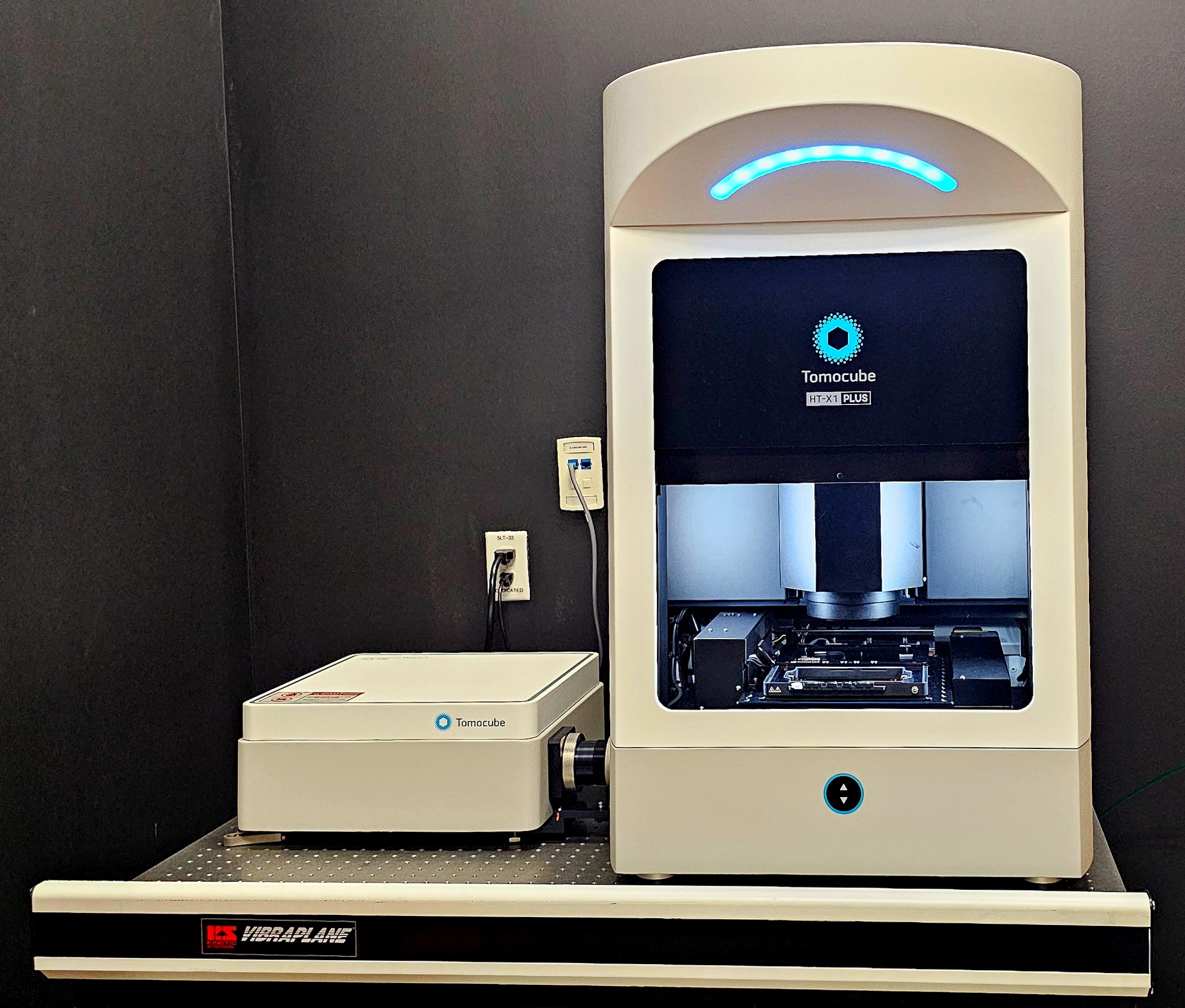
New Tomocube HT-X1 Plus
The QLMC is excited to introduce the Tomocube HT-X1 Plus to the UTSW research community! The Tomocube HT-X1 Plus is an advanced imaging platform that produces high-resolution, three-dimensional images of live cells, organoids, and tissue sections. It’s a new way of visualizing samples, as the core method is optical diffraction tomography, a label-free imaging technique based on measuring how light slows down (phase delay) as it passes through a sample. From these measurements, the instrument computes a 3D map of refractive index, essentially reconstructing the internal structure of cells and tissues without staining. It does so by projecting light at different angles and patterns (via LEDs), and the transmitted light is detected to build up 3D refractive index data. Thus, because it uses low-intensity light, it reduces damage to live cells and allows long-term monitoring. In addition, our new platform integrates a fluorescence module so you can combine detailed structural data (from holotomography) with molecular or biomarker-specific fluorescence signals. Finally, the advanced software uses AI-enhanced algorithms to reconstruct high-quality images quickly and quantitatively (e.g., volume, dry mass, concentration, …). Thus, the Tomocube HT-X1 Plus lets you visualize your samples in a way you’ve never seen before, giving access to data not obtainable via conventional microscopy.
- 10/2025: Demo of the Zeiss LSM910 with Lightfield 4D detector and AiryScan2
Demo of the Zeiss LSM910 confocal with AiryScan2 and Lightfield 4D detector
The QLMC is demoing the latest confocal imaging platform from Zeiss. Hands-on experience is offered 11/03-06 (potentially longer). This new imaging mode from ZEISS is coupled to their proven LSM900 series confocals and enables capturing entire 3D volumes in a single snapshot, of up to 80 volumes per second.
Hands-on demo dates: 10/03-10/06
Demo place: NL5.120E
If you have questions, feel free to contact Lindsey.charley@zeiss.com
- 08/2025: Demo of the Nikon Ji dual camera CSU-W1 with SoRA super-resolution
Demo of the Nikon Ji dual camera CSU-W1 with SoRA super-resolution
The QLMC is demoing the latest confocal imaging platform from Nikon. Hands-on experience is offered 08/12-14 (potentially longer). If you’d like to obtain confocal images (standard or super-resolution) of highest quality, or just have a look at the latest technology, feel free to sign up: System:
Nikon Eclipse Ji inverted microscope
Dual camera CSU-W1 SoRA spinning disc
Back illuminated Kinetix 22 camera
Compact 7-line excitation source Lumencor Ziva 405/446/488/518/577/639/748
Silicone immersion lenses
stage-top incubator for live-cell experiments.Hands-on demo dates: 08/10 - 08/14
Demo place: NL5.120E
If you have questions, feel free to contact Marcel Mettlen (marcel.mettlen@utsouthwestern.edu).
- 02/2025: ZOYC (Zeiss on your campus) event
ZOYC (Zeiss on your campus) event
The QLMC is happy to host a ZOYC (Zeiss on your campus) event. Zeiss will present in a virtual seminar the capabilities of their LSM900 microscope, followed by a hands-on demo.
Webinar: 02/07, 12:00-1:00 PM
Hands-on demo dates: 02/10 - 02/14
Demo place: NL5.120S
If you have questions, feel free to contact Marcel Mettlen (marcel.mettlen@utsouthwestern.edu).
- 12/2024: Meet the ECHO spinning disk confocal
Meet the ECHO spinning disk confocal
Are you interested in a very easy-to-use, relatively inexpensive yet powerful spinning disk confocal? If so, please plan on attending QLMC’s demo days of the ECHO spinning disk confocal:
Hands-on demo dates: 12/10 - 12/15
Demo place: NL5.120S
For more information visit ECHO’s website: ECHO spinning disk
If you have questions, feel free to contact Kevin Lam (klam@discover-echo.com).
- 06/2024: Demo of the Nikon Eclipse Ji with AR-R NSPARC point scanning confocal
Demo of the Nikon Eclipse Ji with AR-R NSPARC point scanning confocal
The QLMC is excited to demo Nikon’s latest product: the versatile Eclipse Ji point scanning confocal with Ax R NSPARC detector.
Demo Dates: 06/06 - 06/15
Place: NL5.120E
For more information visit Nikon’s website: Eclipse Ji
If you have questions, feel free to contact Marcel Mettlen (marcel.mettlen@utsouthwestern.edu).
- 06/2024: Demo of the Tomocube HT-X1
Demo of the Tomocube HT-X1
The QLMC is excited to host a webinar about holographic label-free imaging – a quite powerful and novel way to image certain biological processes. The webinar is presented by Tomocube. The presentation will be followed by a hands-on demo of the HT-X1.
Webinar: 08/13, 12:00-1:00 PM
Hands-on demo dates: 08/20 - 08/29
Demo place: NL5.120S
For more information visit Tomocube’s website: Tomocube
If you have questions, feel free to contact Marcel Mettlen (marcel.mettlen@utsouthwestern.edu).
- 01/2024: New, fully loaded Zeiss LSM980
The Quantitative Light Microscopy Core is adding another high-end system to its microscope suite: a fully specced LSM980. This state-of-the-art setup will meet all your fluorescent imaging needs. Equipped with 8 laser lines (405, 445, 488, 514, 561, 594, 639 and 730nm), it allows for multi-color imaging. Combined with the 32-channel spectral detector, the system can easily handle your multiplexed samples. In addition, the system comes with Zeiss’ proprietary AI sample finder, greatly facilitating and accelerating the acquisition of large tissue sections. The Airyscan2 module allows for fast laser scanning confocal imaging, which is supported by a high-speed piezo z-stage. Alternatively, the Airyscan2 module allows for easy-to-implement super-resolution data acquisition, effectively doubling the optical resolution (down to 120nm). The microscope is located in NL5.120LA.
- 12/2023: New, dual-camera spinning disk confocal
The Quantitative Light Microscopy Core is updating its microscope suite: The heavily used Nikon CSU-W1 SoRa spinning disk confocal finally gets some relief: a new Nikon CSU-W1 spinning disk confocal with 2 cameras. Closely matching the configuration of the “old” system, the new spinning disk does not have the FRAP/SoRa modules, but comes with an additional 594nm laser line and 2 cameras for true simultaneous channel acquisition. The microscope is located in NL5.120R.
- 08/2023: New widefield microscope
The Quantitative Light microscopy core is updating its microscope suite by adding a widefield microscope for epifluorescence and histological stains (color camera). The system is fully motorized for xyz scans. Furthermore, the full enclosure incubator and perfect focus system allows for long-term acquisition of live cells. The microscope is located in NL5.120R.
- 01/2023: 2023 Graduate course "Multiscale Microscopy for Biomedical Research”
2023 graduate course "Multiscale Microscopy for Biomedical Research"
Feel free to follow our "multiscale microscopy for biomedical research" graduate course!
Dates and time: 01/11 - 05/06; Wednesdays and Fridays from 1:30 to 3pm.
Place: Contact Kate Phelps (kate.phelps@utsouthwestern.edu) for more information.
Hands-on labs and demos are limited to registered graduate students. Auditors will be accommodated if space allows. Contact Kate Phelps (kate.phelps@utsouthwestern.edu) to sign up for the waiting list.
- 12/2022: Demo of the Andor Dragonfly spinning-disk confocal and Imaris workshop
Demo of the Andor Dragonfly spinning-disk confocal and Imaris workshop
We are excited to demo Andor's flagship spinning-disk confocal microscope this January: the DragonFly 600. The Dragonfly is an advanced multi-modal imaging system, capable of fast and gentle confocal, widefield, TIRF, and nano-scale super-resolution microscopy.
Demo Dates: 01/09 - 01/13
Place: TBD
Webinar: 01/09 at 12:00pm in NL6.125. Light refreshments will be provided.
For more information visit Andor's website: DragonFly600
If you have questions, feel free to contact Marcel Mettlen (marcel.mettlen@utsouthwestern.edu) or Ryan Robinson (Email).
- 03/2022: Demo of the Zeiss LSM980 with AiryScan2
Demo the Zeiss LSM980 with Airyscan2!
Webinar: 03/11 at 12:00pm
For more information and registration, please visit: Zeiss LSM980
Demo Dates: 03/21 - 03/30
Place: NL5.120R
Announcing the 2022 ImageJ/Fiji Workshop
Dates: May 31, June 7, June 14
Time: 9:00am to noon
Room: D1.502
Instructors: Marcel Mettlen (Director, QLMC) and Kate Luby-Phelps (Director, EMCF)May 31 - A Basic Introduction to Digital Imaging & Image Analysis with ImageJ/Fiji
June 7 - Using ImageJ/Fiji for Specific Applications
June 14 - An Introduction to Scripting/Macro WritingThis workshop is open to all investigators at UT Southwestern. There is no need to RSVP, just show up. Please bring your laptop and have Fiji installed before you come. Links to the downloads with installation instructions are here: https://imagej.net/Fiji/Downloads
Possibly adding new capabilites to the QLMC
The Livecyte is a microscope system able to capture a large amount of high-content images of even the most sensitive cells. With its integrated AI software, the Livecyte monitors and characterizes cell proliferation, cell lineages, motility, morphology and more, from 96 wells to single cells. Currently, no device on campus compares to this system in terms of data quality and amount of information extracted. Below are just 2 examples:
To gage interest, please complete the follwing short poll: Livecyte
Thank you!
- 02/2022: Demo the Evident Scientific FV3000 laser scanning microscope
Demo the Evident Scientific FV3000 laser scanning microscope!
Virtual informational workshop: 02/24 at 12:00pm
Please contact Marcel Mettlen (Marcel.Mettlen@utsouthwestern.edu) for zoom link.
Demo Dates: 03/03 - 03/10
Place: NL5.120R - 01/2022: Graduate course and our photo gallery went live
2022 graduate course "Multiscale Microscopy for Biomedical Research"
Feel free to follow our "multiscale microscopy for biomedical research" graduate course!
Dates and time: 01/10 - 05/06; Mondays and Fridays from 1:30 to 3pm.
Place: Contact Kate Phelps (kate.phelps@utsouthwestern.edu) to obtain the zoom link
Hands-on labs and demos are limited to registered graduate students. Auditors will be accommodated if space allows. Contact Kate Phelps (kate.phelps@utsouthwestern.edu) to sign up for the waiting list.
Call for images to be featured on our photo gallery
The QLMC would like to highlight your research and is looking for images to be featured in our website's photo gallery. If you have data that was acquired on one of our microscopes and you’d like to contribute to this gallery, please submit your image(s) with a quick description (microscope, magnification, sample type, fluorescent probes, …. ) to Marcel Mettlen (Email). We are happy to link this image to your lab's website or publication (if applicable) to create some buzz. Looking forward seeing your beautiful/colorful/amazing/funny images! Thank you.
- 12/2021: LCIF becomes the Quantitative Light Microscopy Core
To better encompass the use of our microscopes and services we provide, the core facility has adopted a new name: Quantitative Light Microscopy Core (QLMC). Although the name changes, our mission remains the same!
- 09/2021: New LCIF Director
After 17 years as director of the LCIF, Dr. Kate Luby-Phelps has decided to reduce her workload by turning the facility over to Dr. Marcel Mettlen (mail:marcel.mettlen@utsouthwestern.edu). Dr. Mettlen has more than fifteen years of light microscopy experience and has been learning the ways of the LCIF since January 2020. Dr. Luby-Phelps will continue as director of the Electron Microscopy Core Facility and will remain associated with the LCIF as a staff member.
Unaffected by this change in leadership, the LCIF will continue to offer affordable access to a variety of state-of-the-art optical microscopes. Imaging modalities include laser-scanning and spinning-disk confocal, multiphoton, wide-field deconvolution, TIRF, and more. In addition, the LCIF offers help and advice with sample preparation, image quantification, automation of data analysis and basic microscope maintenance.
New widefield epi-fluorescence microscope
The Live Cell Image Facility is adding a new microscope to its suite: inverted Nikon Eclipse Ti widefield epi-fluorescence microscope.
- 11/2020: New spinning disk confocal
The Live Cell Image Facility is updating its microscope suite: the well-used Andor spinning disk confocal has been replaced with a new Nikon CSU-W1 SoRa spinning disk confocal. The microscope is located in NL5.120R.
supported by NIH 1S10OD028630-01