News
QLMC news and events
Select a news item to view its details. The newest announcements appear first.
- May 2026: New Leica Stellaris FALCON 8
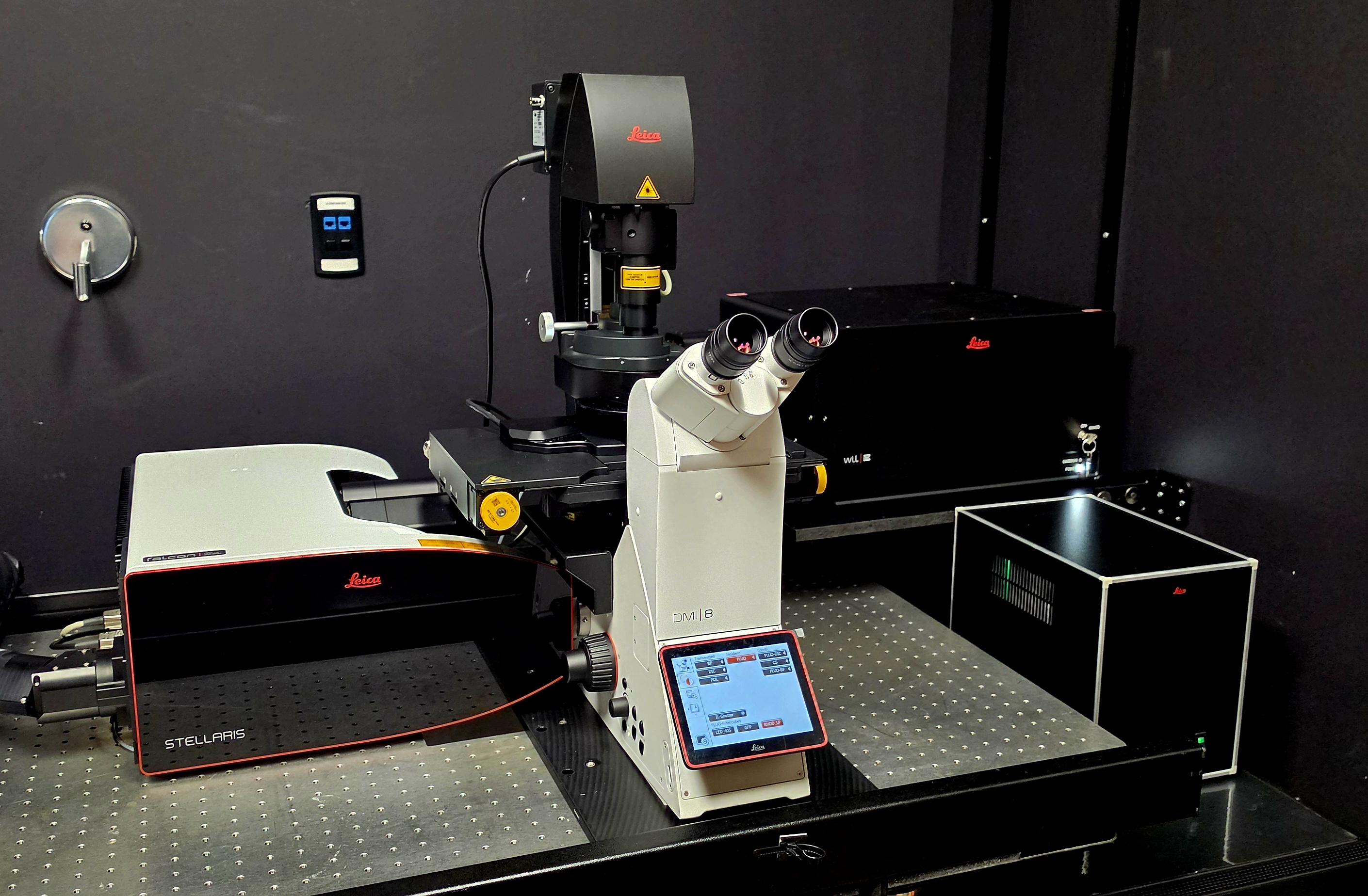
Introducing the Leica Stellaris FALCON 8
The QLMC is excited to introduce the Leica Stellaris FALCON 8 to the UT Southwestern research community. This point-scanning confocal system combines a tunable White Light Laser with sensitive prism-based spectral detection and computer-controlled detection bandwidths. Its broad excitation and detection ranges support highly specific imaging of a wide variety of fluorescent probes.
TauSense adds fluorescence-lifetime information to improve contrast, image quality, and separation of spectrally overlapping fluorophores. The tandem scanning head provides high axial resolution and can switch between resonant and nonresonant scanning. FALCON enables fluorescence lifetime imaging microscopy (FLIM) at confocal speed and integrates directly with LAS X. Analysis options include exponential fitting, phasor analysis, pattern fitting, and FLIM-FRET analysis.
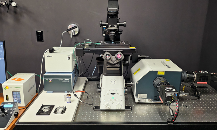
- April 2026: Nikon CSU-W1 dual-camera system moves to South Campus
Nikon CSU-W1 dual-camera system moves to South Campus
The QLMC previously operated only one Zeiss LSM 880 with Airyscan on South Campus. Researchers requiring high temporal resolution therefore had to transport samples to North Campus to use the spinning-disk confocals. At the same time, all available microscope rooms on North Campus were occupied, leaving no space for the new Leica Stellaris FALCON 8 with TauSense.
To expand high-speed imaging access on South Campus and create space for the new Leica system, the Nikon CSU-W1 dual-camera spinning-disk confocal has moved to the newly renovated room K1.228.
- January 2026: New Tomocube HT-X1 Plus
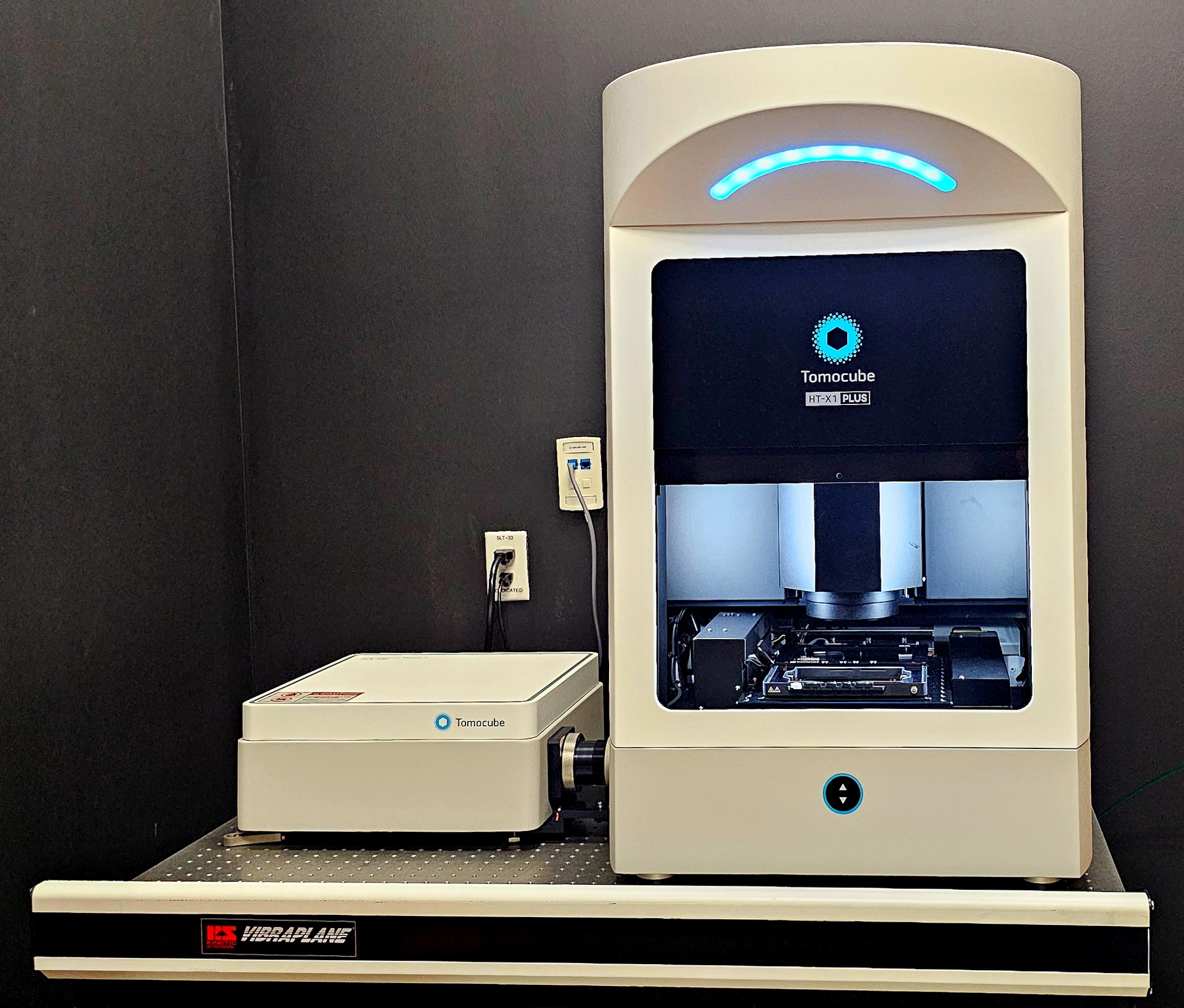
Introducing the Tomocube HT-X1 Plus
The QLMC is excited to introduce the Tomocube HT-X1 Plus to the UT Southwestern research community. This advanced platform produces high-resolution, three-dimensional images of live cells, organoids, and tissue sections using optical diffraction tomography.
This label-free method measures phase delays as light passes through a sample and reconstructs a three-dimensional refractive-index map. Because the system uses low-intensity illumination, it reduces phototoxicity and supports long-term live-cell monitoring. An integrated fluorescence module combines structural holotomography data with molecularly specific fluorescence signals. AI-enhanced reconstruction software also supports quantitative measurements such as volume, dry mass, and concentration.
- October 2025: Zeiss LSM 910 with Lightfield 4D and Airyscan 2 demonstration
Zeiss LSM 910 demonstration
The QLMC is demonstrating the latest confocal imaging platform from Zeiss. The Lightfield 4D detector, coupled to the proven LSM 900-series confocal platform, can capture complete three-dimensional volumes in a single snapshot at rates of up to 80 volumes per second.
Hands-on demonstration: November 3-6, 2025
Location: NL5.120E
For questions, contact lindsey.charley@zeiss.com.
- August 2025: Nikon Eclipse Ji dual-camera CSU-W1 with SoRA demonstration
Nikon Eclipse Ji dual-camera CSU-W1 with SoRA
The QLMC is demonstrating Nikon's latest confocal imaging platform. Researchers are invited to acquire standard or super-resolution confocal images and explore the following system configuration:
- Nikon Eclipse Ji inverted microscope
- Dual-camera CSU-W1 SoRA spinning-disk unit
- Back-illuminated Kinetix 22 camera
- Lumencor Ziva seven-line excitation source: 405, 446, 488, 518, 577, 639, and 748 nm
- Silicone-immersion objectives
- Stage-top incubator for live-cell experiments
Hands-on demonstration: August 12-14, 2025
Location: NL5.120E
For questions, contact marcel.mettlen@utsouthwestern.edu.
- February 2025: Zeiss on Your Campus event
Zeiss on Your Campus
The QLMC is pleased to host a Zeiss on Your Campus event. Zeiss will present a virtual seminar on the capabilities of the LSM 900 microscope, followed by a hands-on demonstration.
Webinar: February 7, 2025, noon-1 p.m.
Hands-on demonstration: February 10-14, 2025
Location: NL5.120S
For questions, contact marcel.mettlen@utsouthwestern.edu.
- December 2024: ECHO spinning-disk confocal demonstration
Meet the ECHO spinning-disk confocal
Researchers interested in an easy-to-use, relatively inexpensive, and powerful spinning-disk confocal are invited to attend the QLMC demonstration.
Hands-on demonstration: December 10-15, 2024
Location: NL5.120S
Learn more about the ECHO spinning-disk confocal.
For questions, contact klam@discover-echo.com.
- June 2024: Nikon Eclipse Ji with AX R NSPARC demonstration
Nikon Eclipse Ji with AX R NSPARC
The QLMC is excited to demonstrate Nikon's versatile Eclipse Ji point-scanning confocal with the AX R NSPARC detector.
Demonstration: June 6-15, 2024
Location: NL5.120E
Learn more about the Nikon Eclipse Ji.
For questions, contact marcel.mettlen@utsouthwestern.edu.
- June 2024: Tomocube HT-X1 webinar and demonstration
Tomocube HT-X1 label-free imaging
The QLMC is hosting a webinar about holographic label-free imaging, followed by a hands-on demonstration of the Tomocube HT-X1.
Webinar: August 13, 2024, noon-1 p.m.
Hands-on demonstration: August 20-29, 2024
Location: NL5.120S
For questions, contact marcel.mettlen@utsouthwestern.edu.
- January 2024: New Zeiss LSM 980 with Airyscan 2
New fully equipped Zeiss LSM 980
The QLMC has added a fully equipped Zeiss LSM 980 to its microscope suite. Eight laser lines at 405, 445, 488, 514, 561, 594, 639, and 730 nm support multicolor fluorescence imaging. A 32-channel spectral detector provides additional flexibility for multiplexed samples.
Zeiss AI Sample Finder facilitates and accelerates acquisition of large tissue sections. Airyscan 2 supports fast confocal imaging with a high-speed piezoelectric z-stage and easy-to-implement super-resolution acquisition with an optical resolution down to approximately 120 nm. The microscope is located in NL5.120LA.
- December 2023: New dual-camera spinning-disk confocal
New Nikon CSU-W1 dual-camera system
The QLMC has expanded its microscope suite with a second Nikon CSU-W1 spinning-disk confocal. Closely matching the heavily used CSU-W1 SoRA system, the new microscope does not include FRAP or SoRA modules but adds a 594 nm laser and two cameras for simultaneous two-channel acquisition. The microscope is located in NL5.120R.
- August 2023: New Nikon Ti widefield microscope
New widefield microscope
The QLMC has added a fully motorized Nikon Ti widefield microscope for epifluorescence and color imaging of histological stains. Motorized x-, y-, and z-axis control supports automated scanning. A full-enclosure incubator and Perfect Focus System enable long-term live-cell imaging. The microscope is located in NL5.120R.
- January 2023: Graduate course in multiscale microscopy
Multiscale Microscopy for Biomedical Research
Researchers are welcome to audit the 2023 graduate course, Multiscale Microscopy for Biomedical Research.
Schedule: January 11-May 6, 2023; Wednesdays and Fridays, 1:30-3 p.m.
Hands-on laboratories and demonstrations are limited to registered graduate students. Auditors will be accommodated when space permits. Contact kate.phelps@utsouthwestern.edu for additional information or to join the waiting list.
- December 2022: Andor Dragonfly demonstration and Imaris workshop
Andor Dragonfly 600
The QLMC is excited to demonstrate Andor's flagship spinning-disk confocal microscope. The Dragonfly 600 is a multimodal imaging system capable of fast and gentle confocal, widefield, TIRF, and nanoscale super-resolution microscopy.
Demonstration: January 9-13, 2023
Webinar: January 9, 2023, at noon in NL6.125. Light refreshments will be provided.
Demonstration location: To be determined
Learn more about the Andor Dragonfly.
For questions, contact marcel.mettlen@utsouthwestern.edu or r.robinson@andor.com.
- May 2022: ImageJ/Fiji workshop
2022 ImageJ/Fiji Workshop
Dates: May 31, June 7, and June 14, 2022
Time: 9 a.m.-noon
Location: D1.502
Instructors: Marcel Mettlen, Director of the QLMC, and Kate Luby-Phelps, Director of the Electron Microscopy Core Facility- May 31: Basic introduction to digital imaging and image analysis with ImageJ/Fiji
- June 7: Using ImageJ/Fiji for specific applications
- June 14: Introduction to scripting and macro writing
The workshop is open to all UT Southwestern investigators. Registration is not required. Please bring a laptop with Fiji installed. Download Fiji and review the installation instructions.
- May 2022: Evaluating Livecyte capabilities for the QLMC
Potential new QLMC capabilities
Livecyte is a high-content imaging system designed to monitor sensitive cells. Its integrated analysis software characterizes cell proliferation, cell lineages, motility, morphology, and other measurements across scales ranging from 96-well plates to individual cells.
Examples:
To help us gauge interest, please complete the short Livecyte survey.
- March 2022: Zeiss LSM 980 with Airyscan 2 demonstration
Zeiss LSM 980 with Airyscan 2
Webinar: March 11, 2022, at noon
Demonstration: March 21-30, 2022
Location: NL5.120R
- February 2022: Evident Scientific FV3000 demonstration
Evident Scientific FV3000 laser-scanning confocal microscope
Virtual informational workshop: February 24, 2022, at noon
Demonstration: March 3-10, 2022
Location: NL5.120R
Contact marcel.mettlen@utsouthwestern.edu for the Zoom link.
- January 2022: Graduate course and QLMC photo gallery
Multiscale Microscopy for Biomedical Research
Researchers are welcome to audit the 2022 graduate course, Multiscale Microscopy for Biomedical Research.
Schedule: January 10-May 6, 2022; Mondays and Fridays, 1:30-3 p.m.
Hands-on laboratories and demonstrations are limited to registered graduate students. Auditors will be accommodated when space permits. Contact kate.phelps@utsouthwestern.edu for the Zoom link or to join the waiting list.
Submit images to the QLMC photo gallery
The QLMC would like to highlight research performed with our instruments. If you have an image acquired on a QLMC microscope, please send the image and a brief description—including the microscope, magnification, sample type, and fluorescent probes—to marcel.mettlen@utsouthwestern.edu. When applicable, we are happy to link the image to your laboratory's website or publication.
- December 2021: LCIF becomes the Quantitative Light Microscopy Core
To better represent the breadth of our microscopy systems and services, the facility has adopted a new name: the Quantitative Light Microscopy Core (QLMC). Although the name has changed, our mission remains the same.
- September 2021: New LCIF Director
After 17 years as Director of the Live Cell Imaging Facility, Kate Luby-Phelps, Ph.D., has transferred leadership of the facility to Marcel Mettlen, Ph.D. Dr. Mettlen has more than 15 years of light-microscopy experience and has worked with the facility since January 2020. Dr. Luby-Phelps will continue as Director of the Electron Microscopy Core Facility and will remain associated with the LCIF as a staff member.
The LCIF will continue to provide affordable access to state-of-the-art optical microscopes, including laser-scanning and spinning-disk confocal, multiphoton, widefield deconvolution, and TIRF systems. The facility also provides guidance on sample preparation, image quantification, analysis automation, and basic microscope maintenance.
New Nikon Eclipse Ti widefield microscope
The LCIF has also added an inverted Nikon Eclipse Ti widefield epifluorescence microscope.
- November 2020: New Nikon CSU-W1 SoRa spinning-disk confocal
The Live Cell Imaging Facility has replaced the heavily used Andor spinning-disk confocal with a new Nikon CSU-W1 SoRa spinning-disk confocal. The microscope is located in NL5.120R.
Supported by NIH award 1S10OD028630-01.