
Metabolites produced by gut bacteria may protect against fungal infection
UTSW-led study shows that byproduct of fiber digestion can fight off C. albicans, which commonly infects immunocompromised patients
DALLAS – July 13, 2026 – A metabolic byproduct formed when gut bacteria break down dietary fiber appears to protect against dangerous fungal infections common in immunocompromised patients, a study led by UT Southwestern Medical Center researchers shows. The findings, published in Cell Host & Microbe, could lead to new therapies to shield this vulnerable population from Candida albicans, a leading human fungal pathogen.
“Patients undergoing cancer therapy, stem cell transplantation, cellular therapy, or prolonged antibiotic treatment frequently experience disruption of their gut microbiome and are at increased risk for invasive fungal infections. By identifying specific microbiota-derived metabolites associated with fungal control, this work provides a foundation for developing dietary, microbiome-based, or metabolite-based therapeutic approaches to restore colonization resistance in high-risk patients,” said study leader Andrew Koh, M.D., Professor of Pediatrics, in the Harold C. Simmons Comprehensive Cancer Center, and of Microbiology. He is also Division Chief of Pediatric Hematology and Oncology at UT Southwestern.
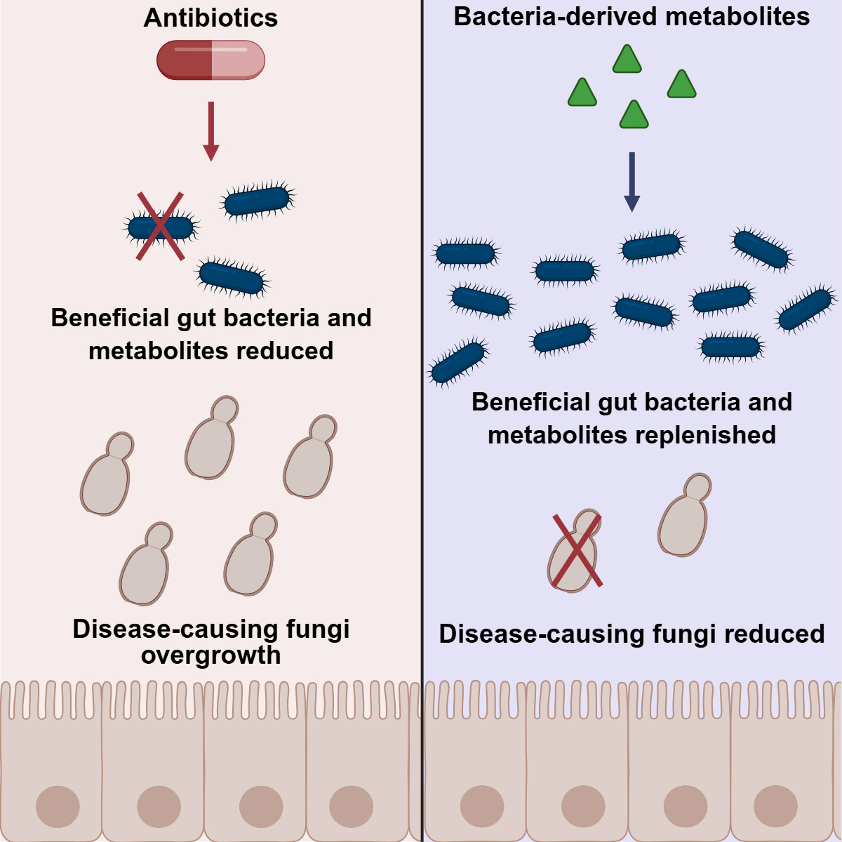
Like virtually all animals, humans have a microbiome – a collection of microorganisms that live on and inside the body, particularly in the intestines. This community of bacteria, fungi, archaea, and viruses typically exists in a harmonious balance, with “good” microbes keeping pathogenic ones in check. However, people whose intestinal microbiome has been disrupted through a variety of immunocompromising conditions can lose this equilibrium. This imbalance can cause C. albicans – a normal part of the microbiome – to become overly prominent, leading to sometimes deadly fungal infections.
In 2015, the Koh Lab found that certain good bacteria appeared to stimulate cells in the intestinal lining to produce a natural antibiotic that provides some protection from C. albicans infections. But whether these bacterial species fight off C. albicans through other mechanisms was unknown.
In the new study, Dr. Koh, his longtime collaborator Lora Hooper, Ph.D., Chair and Professor of Immunology and Professor in the Center for the Genetics of Host Defense and Microbiology, and their colleagues looked for common features of these good bacterial species that keep C. albicans in check. They discovered that all of them produce short-chain fatty acids (SCFAs), lipids primarily made as a byproduct of bacterial digestion of dietary fiber.
When the researchers dosed C. albicans growing in petri dishes with three of these SCFAs – butyric acid, propionic acid, and acetic acid – either alone or together, they found that fungal growth reduced proportionately with increasing SCFA concentrations. Butyric and propionic acids were significantly better at inhibiting C. albicans compared with acetic acid.
Searching for a mechanism, the team compared gene activity in C. albicans cells treated with SCFAs and those left untreated. Results showed that the SCFAs impaired uptake of glucose, a major nutrient for C. albicans; disrupted its ability to digest glucose and other nutrients; and caused the fungal cell interiors to become more acidic. Each of these suppressed pathways led metabolism and growth of C. albicans to slow or shut down, preventing it from overtaking other gut microorganisms.
When the researchers treated mice colonized with C. albicans with SCFAs, they found that this intervention was far more successful in mice with typical intestinal microbiomes than in “germ-free” mice without these bacteria. A closer look showed that the SCFAs encouraged the growth of good bacteria that make these lipids, compounding their anti-fungal effects. When the researchers colonized mouse intestines with bacteria altered to prevent SCFA production, the delivered SCFAs weren’t as effective against C. albicans infections.
The scientists then developed a chemical construct that tied a type of dietary fiber called inulin to propionic acid, which released this SCFA after bacterial digestion in the large intestine, where C. albicans primarily resides. The treatment significantly reduced the amount of this fungus in mouse intestines, suggesting that a similar intervention might be effective in humans as well.
The team plans to continue studying how SCFAs fight C. albicans infections.
A complete list of UTSW authors can be found in the study.
Dr. Koh holds the Grant A. Dove Distinguished Chair for Research in Oncology. He is also a member of the Development and Cancer Research Program at Simmons Cancer Center. Dr. Hooper holds the Jonathan W. Uhr, M.D. Distinguished Chair in Immunology and is a Nancy Cain and Jeffrey A. Marcus Scholar in Medical Research, in Honor of Dr. Bill S. Vowell. She is a member of the Cellular Networks in Cancer Research Program at Simmons Cancer Center.
This research was funded by a grant from the National Institute of Allergy and Infectious Diseases (P01AI179406) and a National Cancer Institute (NCI) Cancer Center Support Grant (P30CA142543).
About UT Southwestern Medical Center
UT Southwestern, one of the nation’s premier academic medical centers, integrates pioneering biomedical research with exceptional clinical care and education. The institution’s faculty members have received six Nobel Prizes and include 28 members of the National Academy of Sciences, 26 members of the National Academy of Medicine, and 14 Howard Hughes Medical Institute Investigators. The full-time faculty of nearly 3,400 is responsible for groundbreaking medical advances and is committed to translating science-driven research quickly to new clinical treatments. UT Southwestern physicians in more than 80 specialties care for more than 143,000 hospitalized patients, attend to more than 470,000 emergency room cases, and oversee nearly 5.3 million outpatient visits a year.