Novel microscopy method at UT Southwestern provides look into future of cell biology
DALLAS – June 28, 2021 – What if a microscope allowed us to explore the 3D microcosm of blood vessels, nerves, and cancer cells instantaneously in virtual reality? What if it could provide views from multiple directions in real time without physically moving the specimen and worked up to 100 times faster than current technology?
UT Southwestern scientists collaborated with colleagues in England and Australia to build and test a novel optical device that converts commonly used microscopes into multiangle projection imaging systems. The invention, described in an article in today’s Nature Methods, could open new avenues in advanced microscopy, the researchers say.
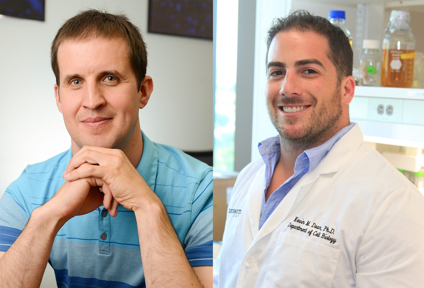
“It is a completely new technology, although the theoretical foundations for it can be found in old computer science literature,” says corresponding author Reto Fiolka, Ph.D. Both he and co-author Kevin Dean, Ph.D., are assistant professors of cell biology and in the Lyda Hill Department of Bioinformatics at UT Southwestern.
“It is as if you are holding the biological specimen with your hand, rotating it, and inspecting it, which is an incredibly intuitive way to interact with a sample. By rapidly imaging the sample from two different perspectives, we can interactively visualize the sample in virtual reality on the fly,” says Dean, director of the UTSW Microscopy Innovation Laboratory, which collaborates with researchers across campus to develop custom instruments that leverage advances in light microscopy.
Currently, acquiring 3D-image information from a microscope requires a data-intensive process, in which hundreds of 2D images of the specimen are assembled into a so-called image stack. To visualize the data, the image stack is then loaded into a graphics software program that performs computations to form two-dimensional projections from different viewing perspectives on a computer screen, the researchers explain.
“Those two steps require a lot of time and may need a very powerful and expensive computer to interact with the data,” Fiolka says.
The team realized it could form projections from multiple angles by optical means, bypassing the need to acquire image stacks and rendering them with a computer. This is achieved by a simple and cost-effective unit consisting of two rotating mirrors that is inserted in front of the camera of the microscope system.
“As a result, we can do all this in real time, without any noticeable delay. Surprisingly, we can look from different angles ‘live’ at our samples without rotating the samples or the microscope,” Fiolka says. “We believe this invention may represent a new paradigm for acquiring 3D information via a fluorescence microscope.”
It also promises incredibly fast imaging. While an entire 3D-image stack may require hundreds of camera frames, the new method requires only one camera exposure.
Initially, the researchers developed the system with two common light-sheet microscopes that require a post-processing step to make sense of the data. That step is called de-skewing and essentially means rearranging the individual images to remove some distortions of the 3D-image stack. The scientists originally sought to perform this de-skewing optically.
While experimenting with the optical de-skewing method, they realized that when they used an incorrect amount of “de-skew,” the projected image seemed to rotate.
“This was the aha! moment. We realized that this could be bigger than just an optical de-skewing method; that the system could work for other kinds of microscopes as well,” Fiolka said.
“This study confirms the concept is more general,” Dean says. “We have now applied it to various microscopes, including light-sheet and spinning disk confocal microscopy.”
Using the new microscope method, they imaged calcium ions carrying signals between nerve cells in a culture dish and looked at the vasculature of a zebrafish embryo. They also rapidly imaged cancer cells in motion and a beating zebrafish heart.
UTSW co-authors include Bo-Jui Chang, Etai Sapoznik, Theresa Pohlkamp, Tamara S. Terrones, Erik S. Welf, Vasanth S. Murali, and Philippe Roudot.
Researchers from the MRC Laboratory of Molecular Biology, Cambridge, United Kingdom; Calico Life Sciences LLC, South San Francisco, California; and the Walter and Eliza Hall Institute of Medical Research and the University of Melbourne, both in Australia, also participated.
The research received support from the Cancer Prevention Research Institute of Texas (RR160057), and the National Institutes of Health (T32CA080621; F32GM117793; K25CA204526, R33CA235254, and R35GM133522). Fiolka has filed a patent for the scan unit and its applications to microscopy. Additional disclosures are in the paper. The researchers made both the data used in the study and the software code available online. Access details are in the study.
Fiolka is a member of the Harold C. Simmons Comprehensive Cancer Center. Visit the Fiolka lab here.
About UT Southwestern Medical Center
UT Southwestern, one of the nation’s premier academic medical centers, integrates pioneering biomedical research with exceptional clinical care and education. The institution’s faculty members have received six Nobel Prizes and include 24 members of the National Academy of Sciences, 25 members of the National Academy of Medicine, and 13 Howard Hughes Medical Institute Investigators. The full-time faculty of more than 3,300 is responsible for groundbreaking medical advances and is committed to translating science-driven research quickly to new clinical treatments. UT Southwestern physicians in more than 80 specialties care for more than 143,000 hospitalized patients, attend to more than 470,000 emergency room cases, and oversee nearly 5.3 million outpatient visits a year.