Neuroimaging
Neuroimaging at the AIRC focuses on studying brain structure, brain networks, functional and vasoreactivity response to physiological modulators, perfusion at rest and in response to tasks, as well as brain metabolism in small animals and humans. Studying various aspects of brain structure, function, and metabolism provides a comprehensive picture of the brain in health and disease.
More details on respective imaging technologies are given in the corresponding pages.
- Ultrahigh Field MRI
- Metabolic MRI
- Hyperpolarized MRI
- NMR Metabolomics
- Molecular Imaging and Probe Development
High-Resolution Mesoscale Anatomical MRI at 7 Tesla (T)
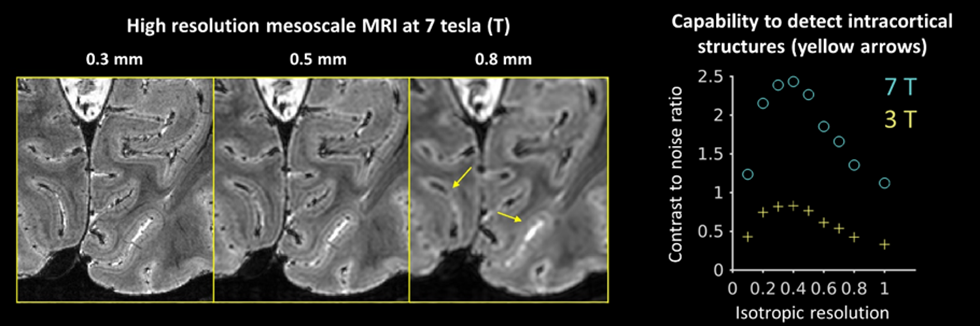
Imaging mesoscale structures in the brain on the order of 0.5 mm can potentially allow us to detect lesions that are not visible using the state-of-the-art clinical imaging methods. The enhanced signal and contrast level provided by ultrahigh field MRI (e.g., 7 T) can help us achieve this goal within a clinically practical scan time of about 10 minutes. As shown in the figure below, cortical layers can be delineated using high resolution MRI at 7 T, but not at lower spatial resolution or lower field strength.
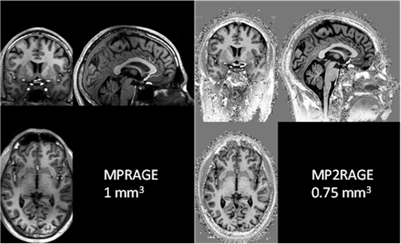
The MP2RAGE scan acquires two whole brain images at different inversion times. By combining these two images, it is possible to create a T1-weighted image without reception field bias and transmit field inhomogeneity. The images below compare the conventional MPRAGE image (1 mm3 isotropic resolution, 7 minutes acquisition time) and the MP2RAGE image (0.75 mm3 isotropic resolution, 6 minutes acquisition time).
Contact PI: Jiaen Liu
Quantitative Neuroimaging
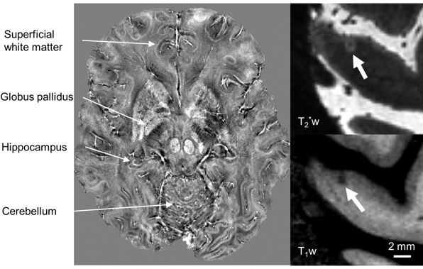
MRI signal is sensitive to various tissue properties in the brain and spinal cord, such as microstructure, iron, myelin, electromagnetic properties, and vascular function. Based on biophysical modeling, quantitative measurement of these parameters can shed light on the disease progress in an early stage for better clinical outcome. The figure below shows quantitative imaging of magnetic susceptibility at 0.3 mm resolution (left), and a cortical lesion of multiple sclerosis at high resolution due to myelin loss.
Contact PI: Jiaen Liu
Imaging Functional Brain Networks
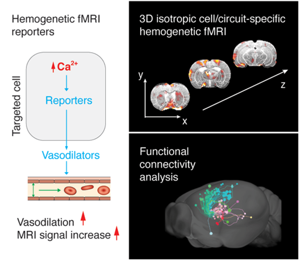
To understand how the brain works, it is crucial to identify the causality of the neural circuits that lead information flow through the whole brain network during certain behavior or task. We develop strategies to visualize and modulate the activity of specific cell populations across the brain using novel whole-brain coverage function MRI. We focus on advancing fMRI techniques from multiple engineering standpoints, including MRI reporter genes, MRI acquisition sequences, and computational models for functional connectivity analysis. Specifically, we aim to implement advanced fMRI techniques with other cutting-edge neural technologies to understand the mechanisms of the reward learning system in rodents, with circuit- and cell-specificity.
Contact PI: Nan Li
Imaging Cerebral Blood Flow
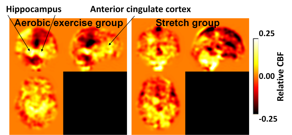
Cerebral blood flow (CBF), the blood vessels’ ability to deliver blood to the brain, can be measured non-invasively using arterial spin labeling (ASL) MRI. CBF provides an index of brain vascular health and a measure reflecting the passive properties of arterial blood vessels to deliver blood to the brain tissue. Our study includes CBF measurements of individuals at high risk for developing Alzheimer’s disease and allows longitudinal monitoring of hippocampal and anterior cingulate CBF with aerobic exercise training.
Contact PI: Binu P. Thomas
Cerebrovascular Reactivity Mapping
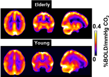
The brain blood vessels’ ability to dilate in response to inhalation of 5% CO2 gas is termed cerebrovascular reactivity (CVR). CVR provides an index of brain vascular health and a measure reflecting the dynamic properties of arterial blood vessels. We investigate CVR responses to CO2 in elderly healthy participants and patients with neurodegenerative diseases or other neurological diseases that affect brain vasculature.
Contact PI: Binu P. Thomas
Metabolic Brain and Spinal Cord Imaging
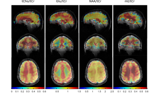
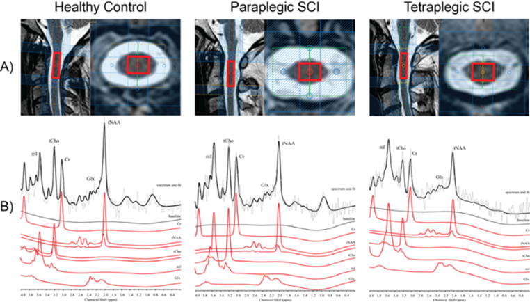
Proton and phosphorous MR spectroscopy and spectroscopic imaging provide rich information about the integrity of neuronal and glial cells, neurotransmission, brain energy metabolism, cell membrane turnover, protein content, and antioxidant levels. Researchers use these techniques to study psychiatric, neurological and neurodegenerative disorders, traumatic injury of the brain and spinal cord, inborn genetic defects in children, as well as brain cancer. The examples below show 1H spectroscopic imaging data of the healthy human brain and single voxel 1H MRS data demonstrating metabolic changes in spinal cord injury.
Contact PI: Anke Henning, Jimin Ren
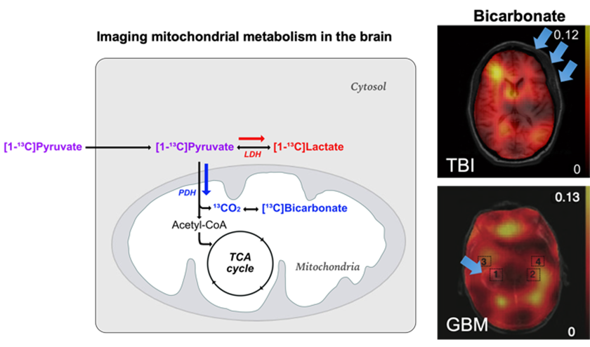
In-Vivo Imaging of Cerebral Metabolism with Hyperpolarized Substrates
The dissolution dynamic nuclear polarization technique (a.k.a. hyperpolarization), in combination with rapid 13C MR spectroscopic imaging, provides a new imaging modality that can assess real-time utilization of substrates and formation of products in-vivo by boosting the signal sensitivity, assessing metabolic pathways of interest, including carbohydrate and branched-chain amino acids metabolism in the brain. With recent advances in clinical translation of hyperpolarized 13C-pyruvate, we have been performing metabolic imaging studies of the human brain in healthy subjects and patients with brain tumors or traumatic brain injury.
Contact PI: Jae Mo Park