Nanomedicine/Drug Delivery for Oncology
Cancer cells undergo pronounced changes in metabolism to enable enhanced proliferative and survival advantages over neighboring non-malignant cells. The research focus in the Corbin Lab investigates strategies that exploit the deviant metabolism of cancer cells (namely the reprogramming of lipid metabolism) for therapeutic purposes.
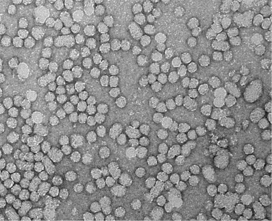
Lipoprotein Nanotechnology
Lipoproteins are naturally occurring core-shell nanostructures. Ranging from 7nm to more than a micron in size, they serve as the main transport vehicles for cholesterol and triacylglycerol in mammalian systems. The low-density lipoprotein (LDL) species interests many cancer researchers because many tumors over-express the LDL receptor (LDLR). Studies in our lab focus on utilizing LDL to deliver anticancer agents to cancer cells.
Formulating conventional drugs into the LDL nanoparticle has produced limited results, as this group adopts an unconventional approach of the reconstituted LDL nanoparticle with bioactive lipids for tumor management/therapy. The natural omega-3 fatty acid, docosahexaenoic acid (DHA), which displays potent anti-inflammatory and anticancer activity, has been the lead active agent in the Corbin Lab LDL nanomedicine program.
Contact PI: Ian Corbin
Image Guided Drug Delivery
The Corbin Lab routinely uses state-of-the–art small animal imaging systems (MRI, micro-MRI, Optical, micro-CT/SPECT/PET) to characterize rodent models of disease. Combining our local expertise in small animal imaging with our nanomedicine technology helps devise unique strategies for sophisticated image-guided drug delivery approaches.
Contact PI: Ian Corbin
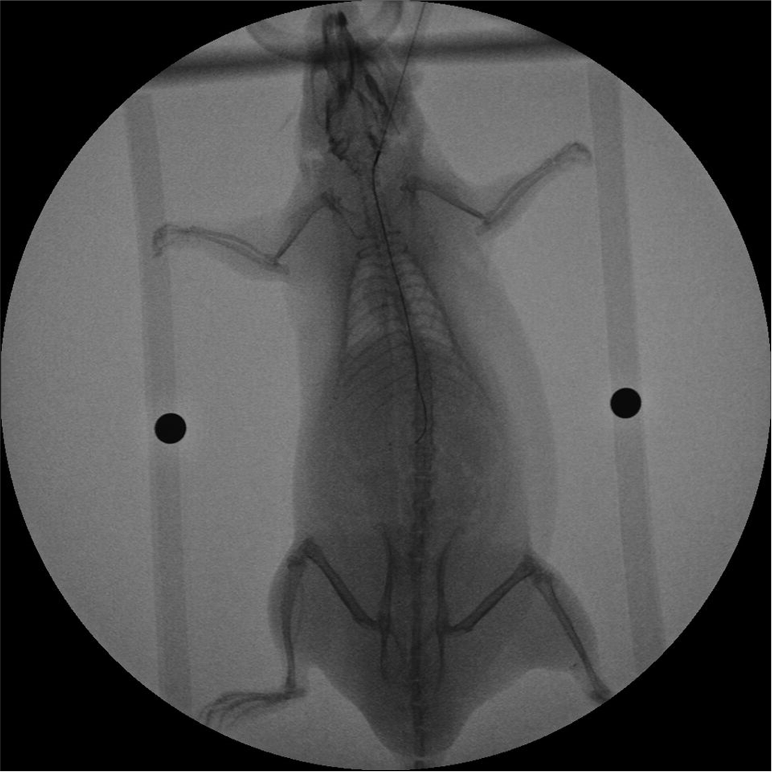
Fluoroscopy
Real time X-ray imaging facilitates loco-regional delivery of nanotherapies to tumors. This technique primarily treats liver tumors. During this procedure, a minimally invasive micro-catheter approach accesses the abdominal and hepatic vasculature. Next, subjects receive X-ray contrast media to delineate the blood vessels feeding the liver and tumor. Upon fluoroscopic confirmation of microcatheter placement, therapeutic agents are seamlessly administered in the local tumor environment.
Contact PI: Ian Corbin
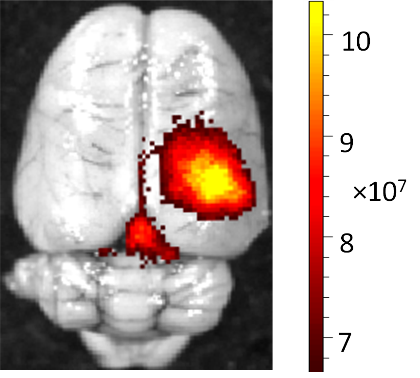
High-Frequency Focused Ultrasound
The application of focused ultrasound (FUS) coupled with IV administration of microbubbles offers a unique method that achieves non-invasive localized opening of the BBB. Low-intensity pulsed FUS exposures delivered to brain regions with circulating intravascular microbubbles cause the microbubbles within the path of the acoustic beam to interact with the endothelial lining, leading to reversible and nondestructive opening of the BBB. MRI guidance enables successful delivery of LDL-based nanomedicine to the brain for the treatment of tumors or other disease processes.
Contact PI: Ian Corbin
Metabolic Hallmarks of Cancer
Metabolic reprogramming is now firmly established as a hallmark of cancer. Tumors share the common phenotype of uncontrolled cell proliferation, which must be sustained by deviant metabolic processes. Successive oncogenic events and the constraints imposed by the tumor microenvironment directly drive the required changes in metabolism.
Contact PI: Ian Corbin
Lipid Reprograming
Development and progression of cancer often link to altered rates of lipid synthesis, mobilization, and metabolism in tumor cells. In collaboration with investigators from UT Southwestern and other institutions, we use mass spectrometry-based lipidomic approaches to interrogate the unique lipid phenotype of malignant tumors.
Contact PI: Ian Corbin