Cardiac and Body MRI
Cardiac and Body MRI research at the AIRC currently focuses on studying function and metabolism of the myocardium, liver, kidney, pancreas, and prostate in small animals and humans. In addition, AIRC faculty take part in collaborative projects with respect to interventional and pediatric cardiac imaging, breast imaging, obesity imaging, as well as low-field MRI.
More details on respective imaging technologies are given in the sections linked in the left menu:
- Metabolic MRI
- Hyperpolarized MRI
- NMR Metabolomics
- Molecular Imaging & Probe Development
- Nanomedicine & Theranostic
Cardiac 1H and 31P MRS
MR spectroscopy in the myocardium can reveal changes in energy metabolism and energy storage. AIRC researchers have expertise with respect to scan protocol development for 1H and 31P single voxel MRS in the human myocardium that include optimal static magnetic field shimming, artefact suppression, and frequency stabilization. Previous clinical trials using 31P MRS revealed changes in the myocardial energy metabolism in acute and subacute Tako-Tsubo cardiomyopathy, hypertrophic cardiomyopathy, diabetes mellitus type 1, as well as heart failure with preserved ejection fraction and partial reversal of the effect by respective treatments. For the first time, we can discriminate between the lipid in the heart muscle cells and the lipid of adipose tissue surrounding the heart by scanning 1H MRS in the human myocardium. This opens the possibility of investigating the impact of energy storage in the metabolic syndrome.
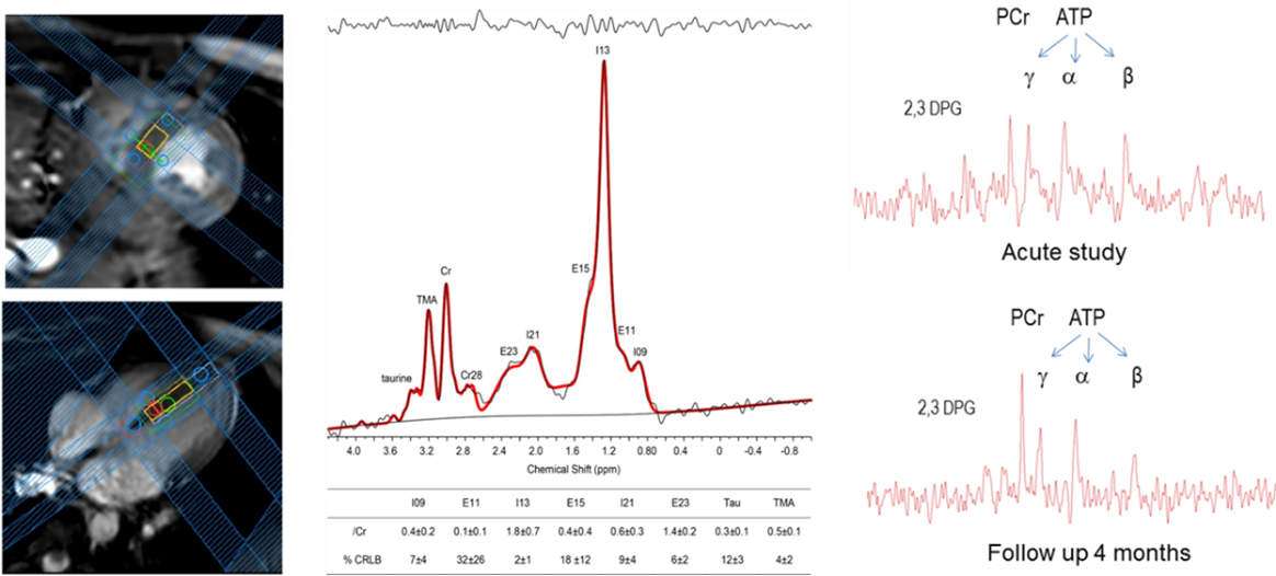
Contact PI: Anke Henning
Cardiac and Kidney MRI
High-resolution, self-gated, cardiac cine imaging can capture the contractile function of the heart. Further post processing of image data can yield heart kinematics including regional myocardial strain. The presence of interstitial fibrosis provides an early biomarker of disease. The extent of fibrosis in the heart and kidney can be quantified by mapping the MR relaxation parameters. Additionally, high resolution T1 and T2 weighted anatomical imaging of the kidney allows clinicians to assess morphological and pathological changes in the kidney.
Impaired renal and cardiac perfusion remains an important marker of disease status, which can be quantified using Arterial Spin Labeling methods without the use of exogenous contrast agents. Black-Blood MR imaging nulls the signal from blood, making it possible to characterize the vessel walls and diagnose inflammatory process of major arteries. Phase contrast MR imaging is used to quantify arterial blood flow. Flow dynamics of aorta and renal arteries not only quantify the stiffness of the artery, but act as a surrogate marker of range of cardiac and renal diseases.
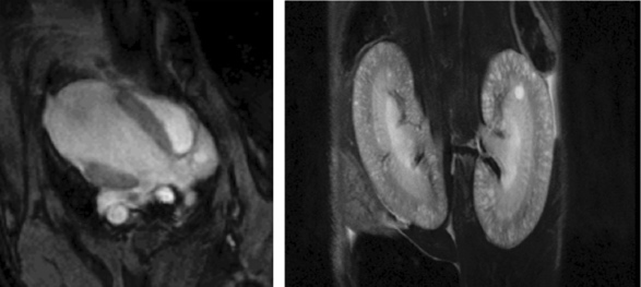
Contact PI: Janaka Wansapura
Monitoring Treatment Response using APT CEST
Amide proton transfer (APT) chemical exchange saturation transfer (CEST) imaging has been introduced as a novel contrast mechanism in the field of molecular imaging. APT CEST MRI can be used to monitor the treatment response of liver cancer bearing rat model to a novel lipoprotein-nanomedicine called LDL-DHA. The LDL-DHA treated tumors displayed a pronounced drop in APT signal, indicative of a significant treatment effect. Histology of the excised tumors confirmed the extensive tumor cell necrosis induced by the LDL-DHA treatment. The changes in endogenous APT signal have been shown to precede changes in conventional MRI and may prove to be a valuable early assessment tool for therapy response.
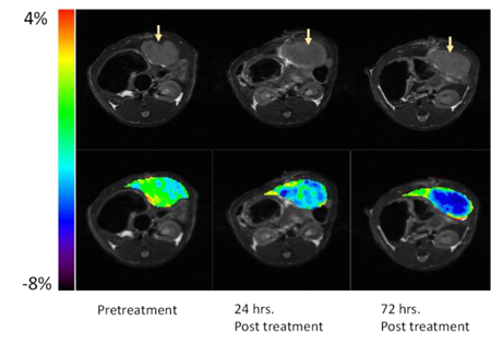
Contact PI: Ian Corbin
Zinc-Responsive MR Contrast Agents
First responder islets initiate an initial burst in insulin secretion in response to a sudden increase in blood glucose concentration while slower responding islets release insulin in over a more prolonged time period. It is important to identify first responder islets because these are thought to be lost during development of type 2 diabetes. Using zinc-responsive MR contrast agents to image release of zinc ions from various tissues in vivo, including the pancreas and prostate.
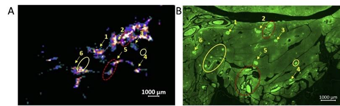
Contact PI: A. Dean Sherry
Cardiac, Hepatic, and Renal Metabolism by Hyperpolarized 13C Substrates
Several probes developed for dynamic nuclear polarization (DNP) that allows real-time assessment of in vivo metabolism in the heart, liver, and kidneys. Hyperpolarized [1-13C]pyruvate can provide new insights into myocardial biology as it is converted by pyruvate dehydrogenase (PDH), a key mitochondrial enzyme for pyruvate oxidation, to [13C]bicarbonate, and by lactate dehydrogenase (LDH) to [1-13C]lactate, the end-product of glycolysis. In addition, gluconeogenic pathways via pyruvate carboxylase (PC) can be identified in the liver and the kidneys (see figure) by detecting PC-specific products such as 13C-labeled malate and aspartate. In addition to rodent studies, we are actively exploring clinical utilities of these technology.
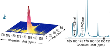
Contact PI: Craig Malloy, Jae Mo Park
Intermediary Metabolism of Kidney and Liver
AIRC researchers study cancer metabolism in patients and intermediary metabolism in the kidney using NMR spectroscopy and stable isotopes. 13C-labeling patterns in lactate from tumors receiving 13C-glucose informed not only glycolysis but also the pentose phosphate pathway. NMR data can also determine liver mitochondrial functions in patients with metabolic syndrome. As shown in the figure, detailed metabolic processes in mitochondria can be measured from an isolated kidney provided with 13C-lactate using isotopomer analysis of multiple metabolites.
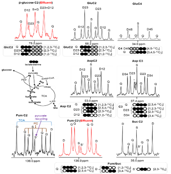
Contact PI: Eunsook Jin, Craig Malloy