Ultrahigh Field MRI
Increasing the sensitivity and spatial resolution of noninvasive medical imaging methods holds great clinical value, potentially providing disease information not available in current clinical practice. Magnetic resonance imaging (MRI) operating at field strength beyond 3 tesla (T), also known as ultrahigh field (UHF) MRI, generates submillimeter spatial resolution, deciphers cortical layer specific responses of the brain to environmental stimuli, and provides previously unavailable metabolite information in a clinically feasible time.
The AIRC hosts the 7T MRI – the highest clinically approved MRI field – and develops cutting-edge enabling technologies in areas including novel contrast, data acquisition and reconstruction, imaging hardware, and calibration methods. We strive for the clinical translation of these promising technologies through collaboration with clinicians on campus and worldwide. While MRI systems with a B0 field strength of 7T and above (i.e. ultrahigh field) provide substantial advantages and potentially improve diagnostic sensitivity and specificity compared to clinical field strength, UHF systems present various technical challenges to overcome before they can be routinely used in the clinic.
Radiofrequency (RF) Coil Design
NE2.511 is home to a state-of-the-art RF engineering laboratory equipped with full-wave simulation servers, precision circuit routers, dielectric probes, network analyzers, 3D printers, and other specialized equipment for design, prototyping and construction. The lab also offers diagnostics and repairs of RF coils, anatomical housings, and biomorphic phantoms. Our RF core staff have the expertise and instrumentation to build workhorse clinical coils, as well as specialized multinuclear coils and novel designs such as high-impedance ultra-flexible coils.
The primary mission of RF lab in AIRC includes:
- Using basic physics research to understand the interaction of RF coils and tissues to improve the sensitivity, transmit efficiency, coverage and safety of RF coils.
- Developing application-specific coils to facilitate research projects at high and ultrahigh field strength.
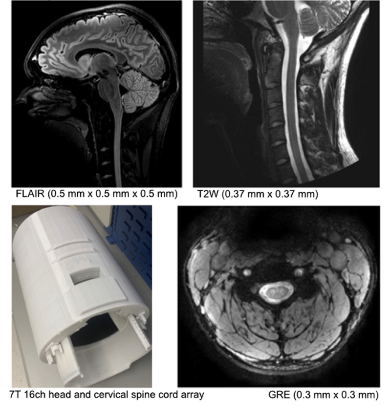
One prime example of our in-house RF coil design is a 7T head and neck coil with extended longitudinal coverage (as compared to the standard commercially available 7T head coil).
Contact PI: Bei Zhang
Parallel Transmission
The shorter electromagnetic wavelength at UHF, which results in inhomogeneity of the radiofrequency (RF) field, can lead to spatially varying flip angles and, thus, spatial variation of the image contrast, signal dropouts, or brightening. The most flexible approach to overcome this issue calls for parallel transmission, which enables much improved control over the spatial and temporal RF field by exploiting the additional degrees of freedom of multiple independent RF transmission channels in combination with spatial encoding via gradients. AIRC researchers explore the concept of universal parallel transmit pulse design with optimized gradient trajectories and applicable to a large flip angle range. This in turn enables the application of this approach to various clinically important MRI sequences.
Contact PI: Anke Henning
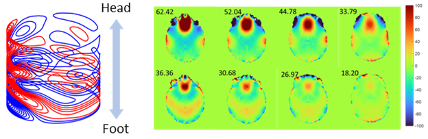
B0 Shimming
B0 field inhomogeneity remains one of the most severe problems in ultrahigh field human MRI. Homogeneity of the static magnetic field (B0) is required to obtain artefact-free images in functional and metabolic MRI scans. Various artifacts can result from poor B0 homogeneity, such as signal dropout, geometric distortion, curved slice profiles, and insufficient peak separation in MR spectroscopy. On one hand, we implemented a versatile stand-alone custom written B0 shimming tool (MATLAB). To complement this software solution, we performed numerical simulations that optimize wire patterns for a dedicated head shim insert. With this hardware brain, B0 shimming can be largely improved in comparison to using the scanner integrated 2nd order spherical harmonics B0 shimming only.
Contact PI: Anke Henning
Applications and Clinical Translation
Emerging clinical applications of ultrahigh field MRI include detecting small lesions and structures in epilepsy, multiple sclerosis, and cartilage degeneration. These mostly utilize high-resolution anatomical imaging. The high spatial resolution and contrast of functional magnetic resonance imaging at ultrahigh fields enables the visualization of complex inter- and even intra-cortical networks. Ultrahigh field MRI allows for highly spatially resolved metabolic imaging and the detection of an enlarged number of metabolites and compounds. These methods are highly valuable in the study of psychiatric and neurodegenerative disorders, cancer in the brain and body, as well as inborn genetic defects.
Examples for current clinical and preclinical ultrahigh field MRI studies:
Contact PIs: Jiaen Liu, Anke Henning, Nan Li