Musculoskeletal MRI
Musculoskeletal MRI research at the AIRC currently focuses on studying the metabolism of the skeletal muscle in humans. More specifically, AIRC faculty participate in collaborative projects relating to the physiological effects of nutrition and exercise. More details on respective imaging technologies are given on this site:
Skeletal Muscle Metabolism by 31P and 1H MRS
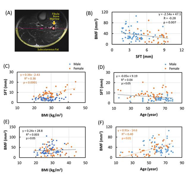
Multiple metabolic imaging studies are implemented and performed at the 3T and 7T whole-body human MRI scanner using 31P MRS in human skeletal muscle as well as fat/water imaging and 1H MRS. For instance, As shown in the figure below, cumulative skeletal muscle proton MRI data at 7T reveled sex differences in subcutaneous fat thickness (SFT) and bone marrow fat (BMF) area and their relationship to BMI and age.
Contact PI: Jimin Ren
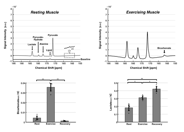
Pyruvate Oxidation Imaged by Hyperpolarized 13C During Exercise
Pyruvate dehydrogenase (PDH) and lactate dehydrogenase remain essential for adenosine triphosphate production in skeletal muscle. At the onset of exercise, PDH dephosphorylation quickly enables the oxidation of glucose and glycogen. However, direct measurement of PDH flux in exercising human muscle is daunting. Lactate and bicarbonate production from hyperpolarized [1-13C]pyruvate in skeletal muscle rapidly reflects the onset and the termination of exercise.
Contact PI: Jae Mo Park