Client Microscopy Facilities
The Histo Pathology Core has microscopy facilities for clients to review and document completed histology slides, as well as to document wet tissues and cell culture preps that augment their histopathology.
Self-Service Microscopy by Reservation
These instruments, located in the Core's Tissue Handling Lab (NB11.204), are offered on a self-service, reservation basis:
- Leica DM2000 Upright Compound Research Photomicroscope
Bright-Field, Dark-Field, Epifluorescence, Polarized Illumination
1.25x, 2.5x, 4x, 10x, 20x, 40x, 63x, 100x (oil) - Zeiss Axiovert 100 Inverted Compound Research Photomicroscope
Bright-Field, Phase-Contrast, DIC, Epifluorescence Illumination
4x, 6.4x, 10x, 16x, 20x, 32x, 40x, 64x - Olympus SZH Upright Stereo Zoom DIssection Photomicroscope
Bright-Field, Dark-Field, Reflected Light, Epifluorescence Illumination, 3.75x - 48x Continuous Zoom
A Jenoptik Gryphax CCD Camera (color, 2700 x 1800 pixels) and computer with image acquisition software is interfaced with each microscope. Image acquisition and camera controller software has variable gain, binning, settings memory, and color/monochrome selectivity.
Additionally, the Core offers a slide scanner:
- Pacific Image Electronics Prime Histo XE Slide Scanner
34mm x 23mm specimen area, 5,000 dpi optical, 25,000 dpi interpolated, 16-bit
Usage of the above listed instruments is free of charge, but closely monitored to ensure good photomicrographs are being obtained, that scope time is being efficiently used, and that the instruments are being well cared for. Clients are asked to limit themselves to reserving two-hour maximum blocks of time, and to schedule an initial training appointment before making full use of the facilities.
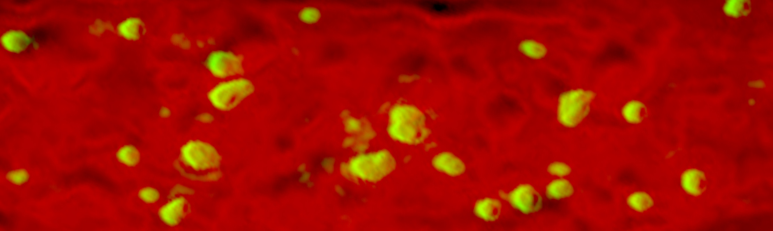
Assisted Fee-for-Service Microscopy
The Core also provides imaging services on a fee-for-service basis for assisted microscopy on the following systems:
- Zeiss Axioplan 2iE Upright Compound Research Photomicroscope
Axiocam CCD (monochrome, 3900 x 3090 pixels), CRI Filterwheel, Improvision Control Suite, Bright-Field, Dark-Field, DIC, Epifluorescence Illumination, Z-Stacking, Deconvolution
1.25x, 2.5x, 4x, 10x, 20x, 40x, 100x (oil) - Nikon E600 Upright Compound Research Photomicroscope
DS-Fi2 (color CCD, 2560 x 1920 pixels), ASI-MS2000, Nikon Elements Control Suite, Bright-Field, Epifluorescence Illumination, Scanning-Stitching
1x, 2x, 4x, 10x, 20x, 40x, 100x (oil) - Zeiss Stemi SV11 Upright Compound Dissection Photomicroscope
Macrofire (color CCD, 2090 x 2090 pixels), LUDL MAC5000, Image Pro-Plus Control Suite, Bright-Field, Dark-Field (conical & incidental angle), Reflected Light, Epifluorescence, Polarized Illumination, Z-Stacking, Extended Depth of Field, 3.78x - 165x Stepped Zoom - Kaiser eVision HF/RTP Gross Photo Stand
Nikkor 60mm Macro (f2.8D), Macrofire (color CCD, 2090 x 2090 pixels), Bright-Field, Reflected Light 200mm2 maximum specimen, 0.2x - 9x Continuous Zoom