Findings could boost efforts to develop chlamydia vaccine
Study co-led by UTSW researchers determines structure of key cell membrane protein in bacterium that causes most common sexually transmitted infection
DALLAS – June 10, 2026 – Scientists at UT Southwestern Medical Center, working with other U.S. researchers, have uncovered the structure of a key cell membrane protein in a bacterial model for Chlamydia trachomatis, the cause of the world’s most common bacterial sexually transmitted infection (STI). The findings, published in Nature Communications, could help guide the design of a vaccine to prevent chlamydia infections, a frequent source of infertility and other health problems.
“Although chlamydia can be successfully treated with antibiotics, a safe and effective chlamydia vaccine could help prevent infections that often go undiagnosed,” said Dominika Borek, Ph.D., Professor of Biophysics and Biochemistry at UT Southwestern. “Many people are asymptomatic yet can still transmit the infection and later develop serious complications. This study provides a structural framework that could aid in the design of more effective vaccine antigens.”

Dr. Borek co-led the study with Luis M. de la Maza, M.D., Ph.D., Distinguished Professor of Pathology at the University of California, Irvine. Scientists at Lawrence Livermore National Laboratory also participated in the research.
According to the Centers for Disease Control and Prevention, an estimated 150 million people worldwide were infected with C. trachomatis in 2023, the last year data were available. This species has several strains called serovars that cause a variety of conditions, including urogenital infections, blindness, and lymphatic disease. Animals can be infected by other chlamydia species, sometimes with devastating results, Dr. Borek said. For example, koalas are endangered due to prevalent Chlamydia pecorum infections.
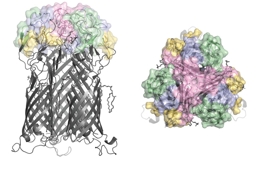
For decades, researchers have tried unsuccessfully to develop a chlamydia vaccine. These efforts have mainly focused on stimulating immunity against the major outer membrane protein (MOMP), found on the exterior of elementary bodies, the bacterium’s infectious stage. Vaccines crafted with denatured or recombinant MOMP – forms lacking the protein’s native three-dimensional structure – haven’t been effective, suggesting its native structure is key to developing immunity against it. However, its three-dimensional structure has been unknown.
To explore this mystery, Dr. Borek, Dr. de la Maza, and their colleagues turned to cryo-electron microscopy (cryo-EM), a technique that can image biomolecules at atomic resolution by freezing them at cryogenic temperatures (lower than -170 degrees Celsius) and examining them with an electron beam. Using UT Southwestern’s Cryo-Electron Microscopy Facility, the researchers imaged MOMP from a chlamydia species that infects mice, called Chlamydia muridarum. They produced images of this protein both alone and bound to a fragment of a mouse anti-chlamydia antibody.
The team’s findings showed that MOMP has an unusual structure unlike any protein seen before, Dr. Borek said, composed of three barrel-shaped units topped with an “antigenic cap” that interacts with antibodies. This cap also displays the protein’s “variable domains,” the part of the protein that diverges in different serotypes. Because MOMP appears to play a role in adhering to host cells – a key step in chlamydia infection – these variable domains influence which types of cells the bacteria can adhere to, determining what types of infection they cause, Dr. Borek explained.
Researchers had previously hypothesized that MOMPs served as molecular pores in elementary bodies’ membranes, she added. However, the team’s findings about its structure show this isn’t possible. The base of the protein acts as a stopper, while the antigenic cap serves as a lid, effectively preventing any molecules from passing through.
The cryo-EM imaging of MOMP’s three-dimensional structure offers a blueprint to guide vaccine development, Dr. Borek said.
Other UTSW researchers who contributed to this study are first author Yirui Guo, Ph.D., M.B.A., Research Scientist, and Zbyszek Otwinowski, Ph.D., Professor of Biophysics and Biochemistry.
This study was funded by a contract with the U.S. Department of Health and Human Services (75N93022C00035) and grants from the National Institute of Allergy and Infectious Diseases (NIAID) and the National Institute of General Medical Sciences (NIGMS), both part of the National Institutes of Health (U19AI144184 and R35GM145365). The Cryo-Electron Microscopy Facility is supported by a grant from the Cancer Prevention and Research Institute of Texas (RP220582).
Dr. Borek and Dr. Otwinowski have a financial interest in Ligo Analytics, which develops software for cryo-electron microscopy.
About UT Southwestern Medical Center
UT Southwestern, one of the nation’s premier academic medical centers, integrates pioneering biomedical research with exceptional clinical care and education. The institution’s faculty members have received six Nobel Prizes and include 27 members of the National Academy of Sciences, 25 members of the National Academy of Medicine, and 13 Howard Hughes Medical Institute Investigators. The full-time faculty of nearly 3,400 is responsible for groundbreaking medical advances and is committed to translating science-driven research quickly to new clinical treatments. UT Southwestern physicians in more than 80 specialties care for more than 143,000 hospitalized patients, attend to more than 470,000 emergency room cases, and oversee nearly 5.3 million outpatient visits a year.