The Titan™ Krios microscope can provide 3D images of biological molecules with up to atomic resolution.
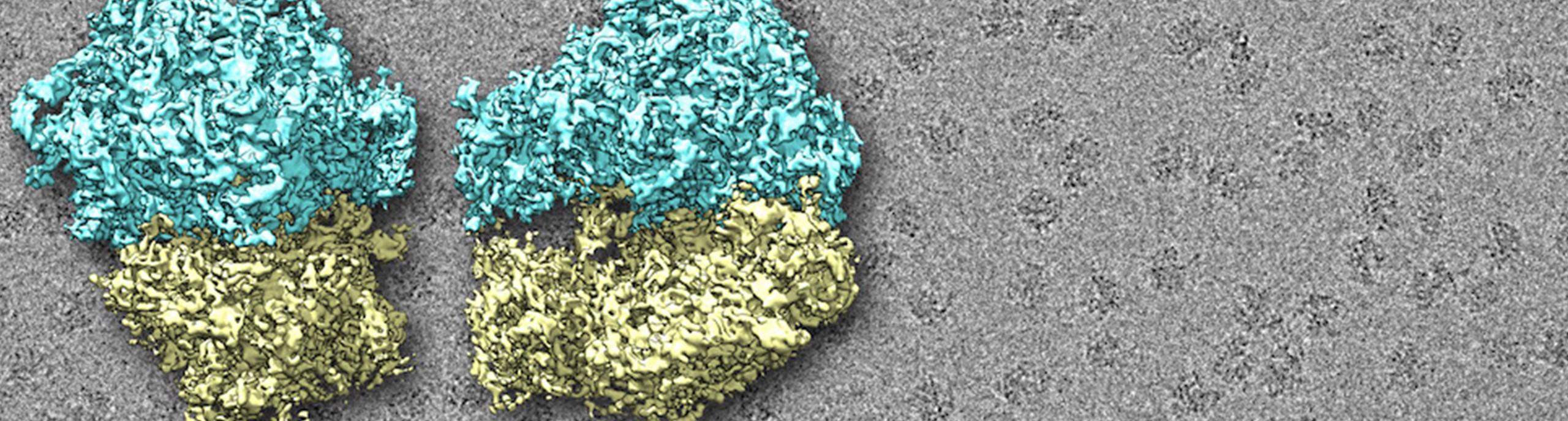
Single-Particle Cryo-EM
Provides 3D reconstructions of macromolecules and their assemblies. Shown: ribosomes.
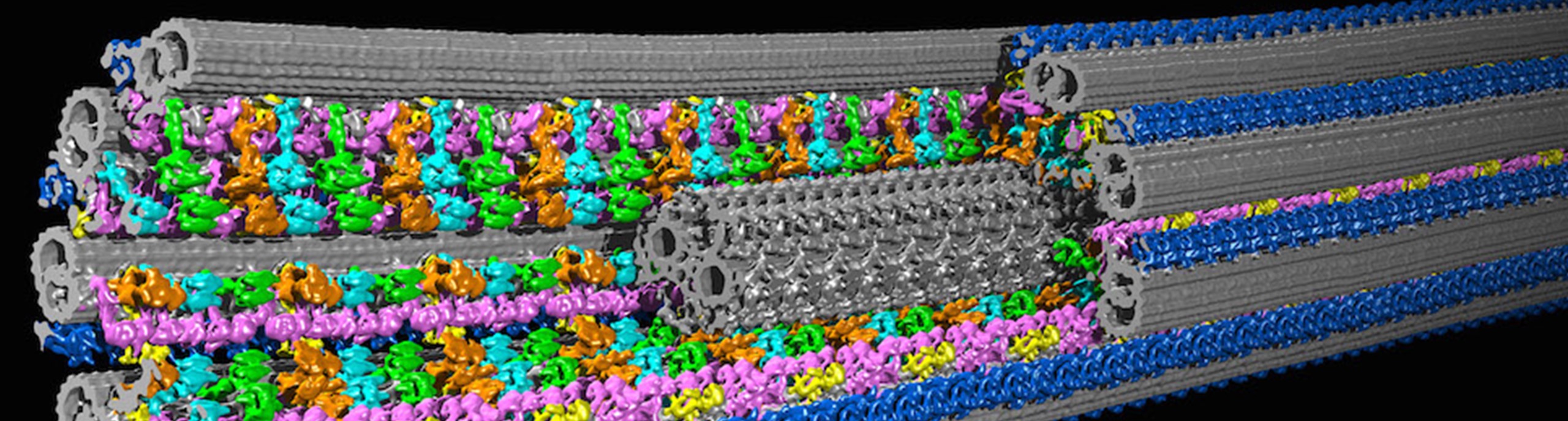
Cryo-Tomography
A new window into the 3D ultra-structure of cells, organelles, and macromolecular complexes. Shown: structure of a cilium.
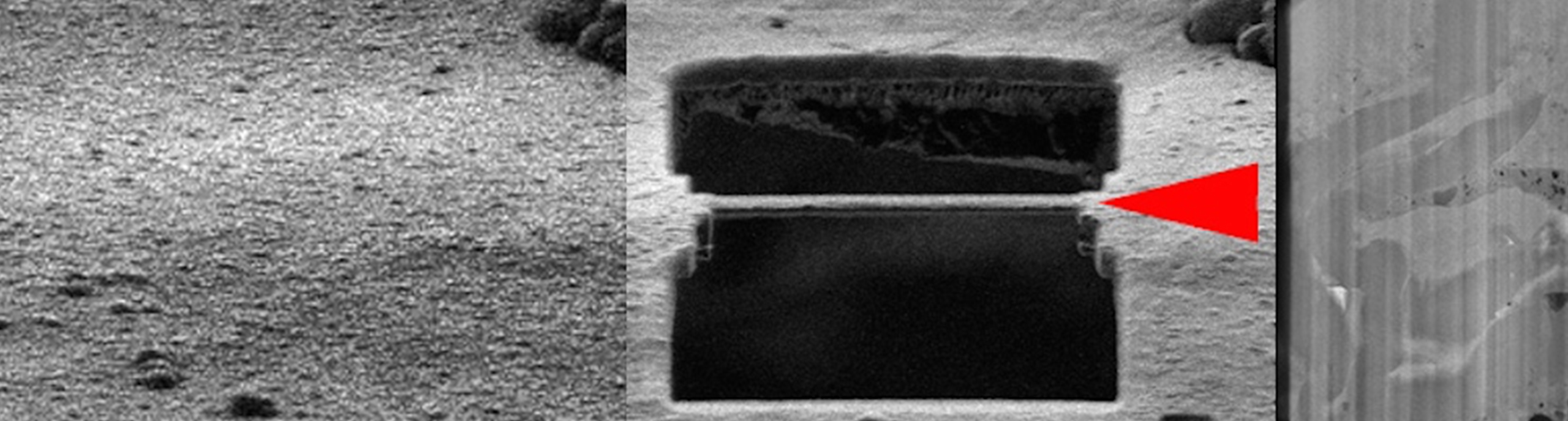
Cryo-FIB Milling
Development of this “nano-sandblaster” will facilitate cryo-tomography deep inside cells.
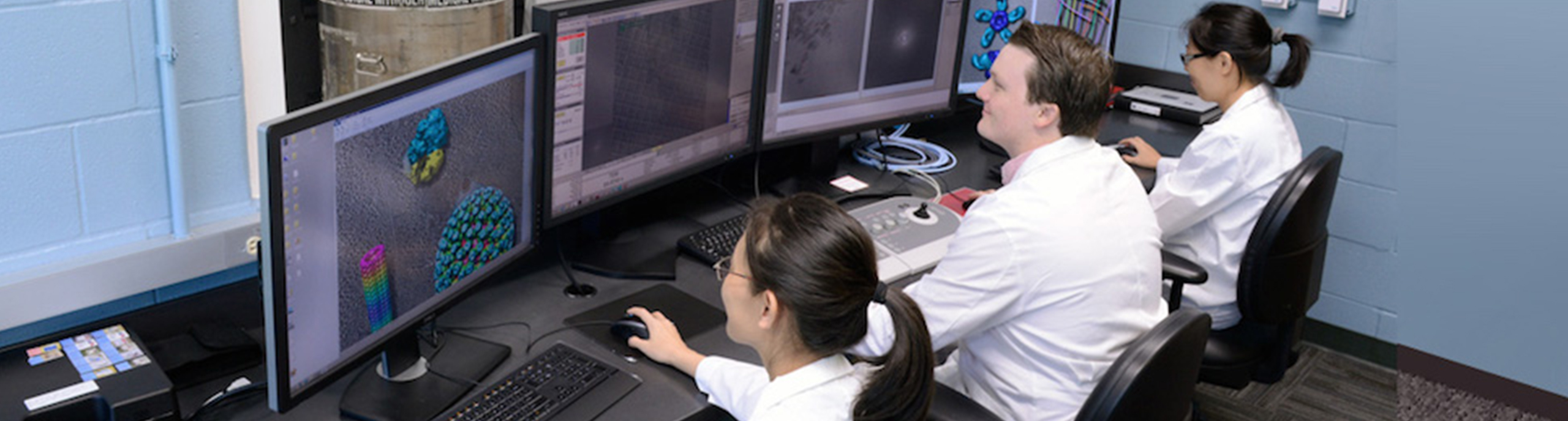
Cryo-Electron Microscopy Facility
This state-of-the-art facility provides access to the 2017 Nobel-prize winning technology of atomic-resolution single particle cryo-electron microcopy (cryo-EM) and cellular cryo-electron tomography to tackle unanswered questions in structural cell biology and to provide a new understanding of the molecular causes of diseases.
UTSW CryoEM Facility Spotlight Daniel J. Stoddard, Ph.D.
 CEMF annex, NL1.180