
Study of living brain tissue reveals how Parkinson’s treatment works
Research co-led by UTSW shows deep brain stimulation strengthens and reshapes ‘building blocks of thoughts and memories,’ cellular systems
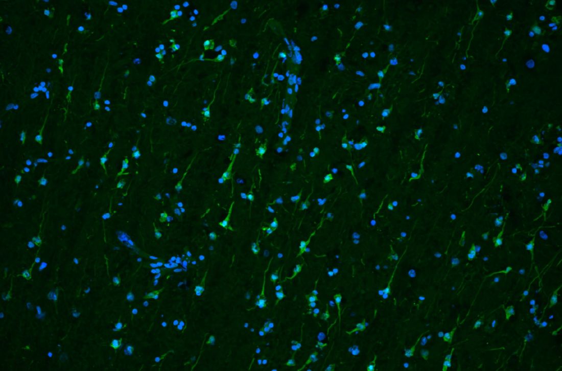
DALLAS – Aug. 10, 2026 – A team co-led by UT Southwestern Medical Center researchers has identified cellular and molecular mechanisms that could explain the effects of deep brain stimulation (DBS), a therapy used to treat movement disorders including Parkinson’s disease and essential tremor. The findings, published in Nature, could lead to new and better ways to apply this treatment.
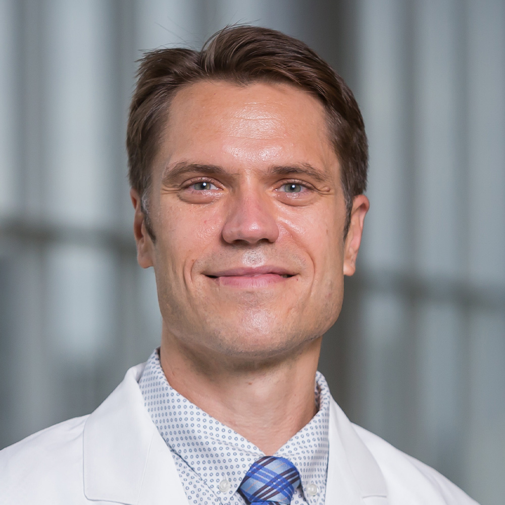
“DBS has transformed the treatment of movement disorders such as Parkinson’s disease, and it’s been explored for neuropsychiatric and cognitive conditions such as obsessive-compulsive disorder, major depression, and dementia. Our study suggests DBS may act as a therapy by modifying both the neuronal building blocks of thoughts and memories and the surrounding cellular systems that help stabilize and reshape them,” said Bradley Lega, M.D., Professor of Neurological Surgery, Neurology, and Psychiatry and an Investigator in the Peter O’Donnell Jr. Brain Institute at UT Southwestern.
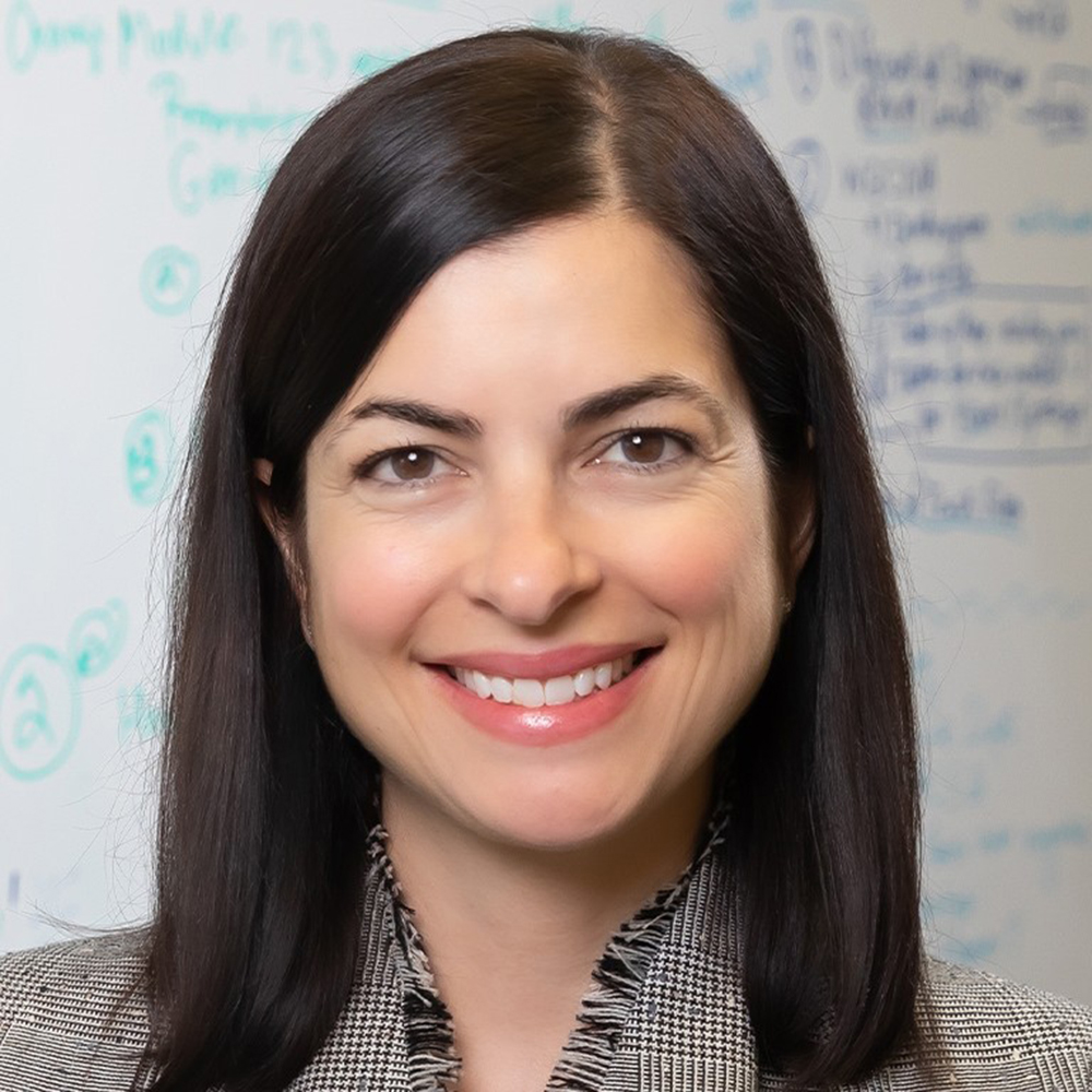
Dr. Lega co-led the study with Genevieve Konopka, Ph.D., Chair of Neurobiology in the David Geffen School of Medicine at the University of California-Los Angeles (UCLA) and a former Professor and Vice Chair of Neuroscience and current Adjunct Professor of Neuroscience at UTSW.
In DBS, electrodes surgically implanted in the brain deliver controlled electrical impulses. Since its inception in the 1960s, researchers have hypothesized this stimulation disrupts pathological patterns of brain activity, allowing neural circuits to rewire and promote healthier electrical signaling between neuronal assemblies, groups of neurons that fire together and are believed to serve as the building blocks of thought and memory.
However, exactly how DBS changes assembly behavior, and the genetic and molecular underpinnings of these changes, has been unknown. Studying this phenomenon has been difficult since lab experiments on brain tissue still in living patients aren’t possible, and the brains of humans differ significantly from animal and organoid models, Dr. Lega explained.
So Dr. Lega – a neurosurgeon who frequently treats patients with epilepsy by removing causative brain tissue – and his colleagues developed a way to keep surgically extracted human brain tissue alive in the lab for study, an approach he said had never been successfully accomplished for the purposes of studying mechanisms of DBS.
Working with tissue donated by 12 patients with epilepsy at UT Southwestern and Parkland Memorial Hospital, Drs. Lega and Konopka and their colleagues placed each sample on a device containing thousands of microscopic electrodes that delivered stimulation to individual cells and recorded their electrical activity. Through these experiments, they identified 19 assemblies and found that stimulating the assemblies for 20 minutes boosted their firing strength. It also increased the assemblies’ plasticity, heightening their ability to add or subtract cells.
When the researchers compared the assemblies’ gene activity before and after electrical stimulation, they found that stimulation appeared to affect the output of genes involved both in neuronal activation and plasticity as well as communication between synapses, the connections between neurons.
Gene activity wasn’t altered only in neurons, Dr. Lega said. The team also found differences in glial cells, which support neurons by regulating their excitability, promoting synaptic communication and remodeling, and controlling the speed and reliability of electrical signals.
Much of this gene activity was mirrored in tissue surgically removed from eight patients with epilepsy just after they underwent DBS, suggesting the lab experiments closely mimicked the effects of this therapy.
The researchers say they plan to continue studying how DBS affects brain cells using larger numbers of samples. They’re grateful to the epilepsy patients who are willing to donate this tissue after it has been removed for clinical purposes.
They are particularly interested in investigating the effects of stimulation protocols similar to those currently used for patients with movement disorders, who typically receive constant stimulation after their electrodes are activated rather than the short, transient stimulation used in this study.
“These results give us the first insights into the genes and specific cell types that are activated with DBS-like stimulation patterns,” Dr. Konopka said. “We believe this knowledge will allow us to develop more targeted and effective therapies for many forms of cognitive issues, especially memory impairment, in the future.”
Other UTSW researchers who contributed to this study are first author Haley Moore, Ph.D., a medical student in UTSW’s Perot Family Scholars Medical Scientist Training Program, and Aswinikumar Kulkarni, Ph.D., Assistant Professor in the O’Donnell Brain Institute and of Neuroscience.
Dr. Lega holds the William Kemp Clark Chair of Neurological Surgery.
This research was funded by the O’Donnell Brain Institute and grants from the National Institutes of Health (NS132443, NS126143, GM148302, and CA138313).
About UT Southwestern Medical Center
UT Southwestern, one of the nation’s premier academic medical centers, integrates pioneering biomedical research with exceptional clinical care and education. The institution’s faculty members have received six Nobel Prizes and include 28 members of the National Academy of Sciences, 26 members of the National Academy of Medicine, and 14 Howard Hughes Medical Institute Investigators. The full-time faculty of nearly 3,400 is responsible for groundbreaking medical advances and is committed to translating science-driven research quickly to new clinical treatments. UT Southwestern physicians in more than 80 specialties care for more than 143,000 hospitalized patients, attend to more than 470,000 emergency room cases, and oversee nearly 5.3 million outpatient visits a year.
About Parkland Health
Parkland Health is one of the largest public hospital systems in the country. Premier services at the state-of-the-art Parkland Memorial Hospital include the Level I Rees-Jones Trauma Center, the only burn center in North Texas verified by the American Burn Association for adult and pediatric patients, and a Level III Neonatal Intensive Care Unit. The system also includes two on-campus outpatient clinics – the Ron J. Anderson, MD Clinic and the Moody Outpatient Center, as well as more than 30 community-based clinics and numerous outreach and education programs. For more information, visit parklandhealth.org.