Stressors damage kidneys by mutating mitochondrial DNA
UTSW study may provide insight into the decline of other organs and tissues in the body
DALLAS – Oct. 14, 2025 – Kidney damage that seemingly heals appears to mutate the DNA in the mitochondria of kidney cells, making the organ less resilient to future stressors and reducing its function over time, a study led by UT Southwestern Medical Center researchers shows. Their findings, published in Science, could lead to new treatments for acute kidney injury (AKI) and chronic kidney disease (CKD) and may explain some facets of aging in other organs and tissues throughout the body.
“We may have stumbled upon a new way to count the wear and tear that cumulatively degrades the health of long-lived cells,” said study leader Samir Parikh, M.D., Professor of Internal Medicine and Pharmacology and Chief of the Division of Nephrology at UT Southwestern, who specializes in AKI and CKD.

Kidney-related conditions are extremely common: CKD affects more than 1 in 7 U.S. adults, and about 20% of hospitalized adults are diagnosed with AKI. One reason is the extraordinary amount of physiological stress that kidneys experience long term, Dr. Parikh said. Because kidneys are continuously filtering blood to maintain its chemistry, they are exposed to high concentrations of electrolytes, environmental toxins, and potentially damaging medicines such as lifesaving cancer chemotherapies. Because kidney cells live a long time with little regenerative capacity, Dr. Parikh and colleagues wondered whether wear and tear gradually degrades kidney function – and if so, what mechanism might be responsible.
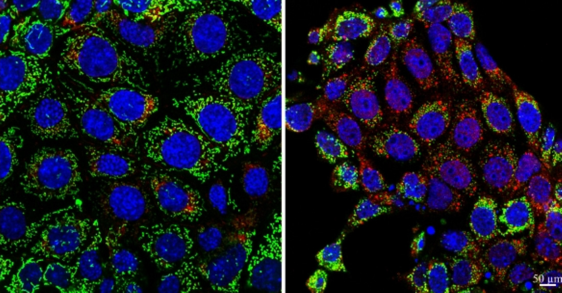
The researchers examined kidney cells from animal models exposed to stressors, such as decreased blood flow to the kidneys or exposure to a damaging chemical. They found these events caused a burst of mutations in the DNA of mitochondria – organelles that act as energy-producing powerhouses – that wasn’t repaired over time, even after the kidneys apparently healed. The researchers found similar mitochondrial DNA mutations in kidney cells exposed to hydrogen peroxide – causing damage known as oxidative stress – and in kidney cells from patients with CKD, suggesting these mutations are a universal signature of physiological stress.
When the researchers used genetic engineering to introduce similar mutations in kidney cells that had never been exposed to these stressors, they found the mitochondria produced significantly less ATP – the energetic molecule that fuels all cellular operations. Kidney cells carrying these mutations were also less resilient when they were exposed to stressors, further declining in function over time.
To tie these findings back to kidney function, the researchers examined mitochondrial DNA from patients with CKD in the UK Biobank, a database containing genetic, health, and lifestyle information from over half a million volunteers. Those with worse kidney function had proportionally more mutations. These results suggest the mutations could signal damage over time, Dr. Parikh said, and predict decline in those whose kidney function was still relatively good. Independently, the burden of mutations predicted the likelihood of future episodes of AKI.
The findings also could explain some general features of aging, he added. Like kidney cells, those that compose the brain, heart, and skeletal muscles are long-lived with little regenerative capacity and perform a large amount of “work” for the body. Consequently, they carry a high number of mitochondria to fuel their activity. These are also tissues that degenerate significantly during aging, with this decline causing many age-related conditions.
If future research shows the mitochondrial DNA of these tissues is also damaged by a lifetime of stress, finding a way to compensate or heal this damage with supplements or by harnessing natural mechanisms that cells use to replace mitochondria could potentially extend both life and health spans, Dr. Parikh said.
Other UTSW researchers who contributed to this study are Vamsidhara Vemireddy, M.D., M.H.A., M.B.A., Manager of Clinical Research Programs for Neurology, and Amanda Clark, Assistant Professor of Pediatrics.
Dr. Parikh holds the Robert Tucker Hayes Distinguished Chair in Nephrology, in Honor of Dr. Floyd C. Rector, Jr., and the Ruth W. and Milton P. Levy, Sr. Chair in Molecular Nephrology.
The study was funded by grants from the National Heart, Lung, and Blood Institute; the National Institute on Aging; and the National Institute of Diabetes and Digestive and Kidney Diseases (R01HL144569, R01AG085753, R01AG027002, U01DK04308, and R01DK095072).
About UT Southwestern Medical Center
UT Southwestern, one of the nation’s premier academic medical centers, integrates pioneering biomedical research with exceptional clinical care and education. The institution’s faculty members have received six Nobel Prizes and include 24 members of the National Academy of Sciences, 25 members of the National Academy of Medicine, and 13 Howard Hughes Medical Institute Investigators. The full-time faculty of more than 3,300 is responsible for groundbreaking medical advances and is committed to translating science-driven research quickly to new clinical treatments. UT Southwestern physicians in more than 80 specialties care for more than 143,000 hospitalized patients, attend to more than 470,000 emergency room cases, and oversee nearly 5.3 million outpatient visits a year.