Underlying cause of Gulf War illness confirmed in UTSW study
Findings could lead to possible treatments for veterans’ debilitating symptoms
DALLAS – Nov. 20, 2025 – Dysfunctional mitochondria, organelles that serve as cellular power generators, appear to cause the symptoms of Gulf War illness (GWI) among tens of thousands of veterans of the Persian Gulf War, UT Southwestern Medical Center scientists confirmed in a new study. The findings, published in Scientific Reports, could lead to effective treatments for this condition that has plagued former soldiers for 30-plus years.
“Our research shows that these veterans don’t have damaged neurons, which would be incurable, but an energy imbalance, which suggests that their disabling symptoms might respond to novel treatments,” said study leader Robert Haley, M.D., Professor of Internal Medicine in the Division of Infectious Diseases and Geographic Medicine and in the Peter O’Donnell Jr. School of Public Health at UT Southwestern. He co-led the study with Sergey Cheshkov, Ph.D., former Assistant Professor of Radiology at UT Southwestern and now Research Scientist/Physicist in the Sammons BrainHealth Imaging Center at The University of Texas at Dallas, and Richard W. Briggs, Ph.D., retired Professor of Radiology at UT Southwestern.
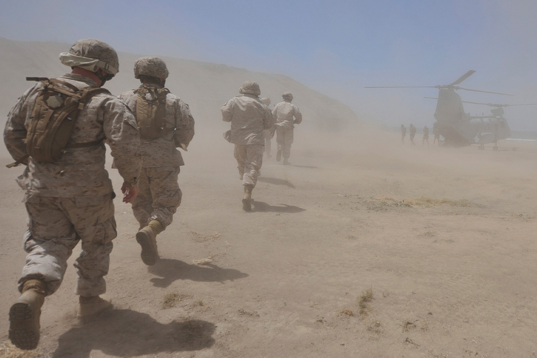
Between 1990 and 1991, about 700,000 U.S. troops leading a multinational coalition were deployed to the Persian Gulf to fight against Iraq. More than 25% of these soldiers returned home with a mysterious collection of chronic symptoms, including fatigue, pain, memory/concentration problems, balance disturbances, exercise intolerance, diarrhea, skin rashes, and depression. A wealth of evidence has tied this condition – now known as GWI – to low-level exposure to sarin gas, a neurotoxic agent often used as a chemical weapon that was released when soldiers bombed Iraqi chemical weapons factories. But how this exposure led to GWI’s symptoms has been a mystery.
For more than three decades, Dr. Haley and his colleagues have searched for answers. One tool they’ve used in their investigations is magnetic resonance spectroscopy (MRS), a brain scanning technique that can quantify chemicals deep in the brain and elsewhere. Starting in 1997, the researchers used this technique to examine the ratio of two chemicals – N-acetylaspartate (NAA) and total creatine (tCr) – in a deep part of the brain called the basal ganglia, which plays a crucial role in coordinating movement, learning, and emotional processing.
Their initial study showed that veterans with GWI had a lower NAA/tCr ratio than those who had also served in the war but didn’t have GWI. However, MRS technology available at the time couldn’t distinguish whether this ratio was due to relatively lower NAA or higher tCr, which have opposing implications: Decreased NAA suggests a process that stems from nerve damage, while increased tCr suggests a process that stems from mitochondrial dysfunction.
In the new study, researchers used state-of-the-art MRS equipment and techniques to measure the NAA/tCr ratio in 39 Persian Gulf War veterans with GWI and 16 without. They found that the lower NAA/tCr ratio in veterans with GWI is due to increased tCr. Because mitochondrial dysfunction in the brain causes chronic neuroinflammation, this finding explains nearly all the symptoms of GWI, Dr. Haley said.
He and his colleagues are currently studying how low-level sarin gas exposure causes mitochondrial dysfunction. This research may eventually lead to treatments that calm this chronic neuroinflammation, bringing relief to those with GWI.
Dr. Haley, a Distinguished Teaching Professor, holds the U.S. Armed Forces Veterans Distinguished Chair for Medical Research, Honoring Robert Haley, M.D., and America’s Gulf War Veterans.
This study was funded by IDIQ contract VA549-P-0027, awarded and administered by the Dallas Veterans Affairs Medical Center, and grants from the U.S. Army Medical Research and Materiel Command (DAMD17-01-1-0741); the Office of the Assistant Secretary of Defense for Health Affairs, through the Gulf War Illness Research Program (W81XWH-16-1-0740); and the North and Central Texas Clinical and Translational Science Initiative, from the National Center for Research Resources of the National Institutes of Health (NIH) and NIH Roadmap for Medical Research (UL1RR024982). The content does not necessarily reflect the position or the policy of the federal government or the sponsoring agencies, and no official endorsement should be inferred.
About UT Southwestern Medical Center
UT Southwestern, one of the nation’s premier academic medical centers, integrates pioneering biomedical research with exceptional clinical care and education. The institution’s faculty members have received six Nobel Prizes and include 24 members of the National Academy of Sciences, 25 members of the National Academy of Medicine, and 13 Howard Hughes Medical Institute Investigators. The full-time faculty of more than 3,300 is responsible for groundbreaking medical advances and is committed to translating science-driven research quickly to new clinical treatments. UT Southwestern physicians in more than 80 specialties care for more than 143,000 hospitalized patients, attend to more than 470,000 emergency room cases, and oversee nearly 5.3 million outpatient visits a year.