Circadian gene may be a key to humans’ unique cognitive abilities
CLOCK gene, discovered by UTSW researcher, influences brain structure and function in addition to regulating circadian rhythm
DALLAS – July 31, 2025 – The CLOCK gene, which serves as a master controller of circadian rhythms, may play a key role in the extraordinary cognitive abilities of humans as well as neuropsychiatric disorders that afflict them, UT Southwestern Medical Center researchers report. Their findings, published in Nature Neuroscience, shed light on human evolution and could lead to new treatments for seasonal affective disorder, depression, schizophrenia, and other conditions.
“Our study suggests the human CLOCK gene, a core circadian regulator, evolved new spatiotemporal expression patterns that contribute to complex brain architecture and advanced cognition,” said Joseph Takahashi, Ph.D., Chair and Professor of Neuroscience at UT Southwestern. Dr. Takahashi co-led the study with Genevieve Konopka, Ph.D., former Professor of Neuroscience at UTSW and now Chair of Neurobiology in the David Geffen School of Medicine at the University of California-Los Angeles, and first author Yuxiang Liu, Ph.D., former Instructor of Neuroscience at UTSW.

Dr. Takahashi discovered the mouse version of CLOCK, known as Clock, in 1997 and the human CLOCK gene in 1999. The gene produces a transcription factor, a protein that controls the activity of other genes. While many genes controlled by CLOCK regulate the rhythmic cycling of biological functions central to the body’s 24-hour circadian clock, other affected genes don’t appear to have a circadian role, Dr. Takahashi explained.
In 2012, Dr. Konopka discovered that human CLOCK has increased expression in the cerebral cortex – an area of the brain that is thought to be key to advanced cognition and has been linked to a variety of neuropsychiatric and neurologic conditions. These discoveries suggested CLOCK has “extra-circadian” functions unique to humans, although those have been unclear.
Dr. Konopka added that the traditional means of studying the functions of genes – either in cells growing in petri dishes or using analogous versions in animals such as mice – don’t resolve how CLOCK affects brain cells in their native environment or behavior in a living organism. Thus, she and her colleagues needed to take a different approach to better understand CLOCK’s functions.

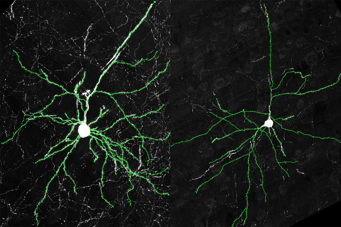
In the new study, the researchers developed “humanized” mice, replacing their native Clock gene with the human CLOCK gene during embryonic development. After the mice became adults, Drs. Konopka, Takahashi, Liu, and their colleagues compared them to mice that carried extra copies of the mouse Clock gene and “wild-type” mice that weren’t genetically altered.
The team found that brains of mice carrying multiple copies of the human CLOCK gene expressed it in a pattern similar to humans, with more of the CLOCK protein made in the cerebral cortex relative to other areas of the brain. This region also became denser, with more cells than the cerebral cortexes of mice carrying extra copies of mouse Clock or wild-type mice.
A closer look showed the excitatory neurons (cells that transmit electrical signals) in the cerebral cortexes of humanized mice grew more dendrites and spines compared with those in the other mice – anatomy that allows for greater connectivity with other neurons. These cells displayed an opposite presentation to human neurons growing in petri dishes in which CLOCK had been deleted; those cells grew fewer dendrites and spines compared with unaltered human neurons and made fewer connections to other neurons.

To determine whether the differences seen in the humanized mice affected their behavior, the researchers had the animals complete a complex cognitive task that required them to learn a changing set of associations to receive a food reward. Mice carrying the CLOCK gene performed significantly better than the other two groups, suggesting enhanced cognitive abilities.
When the researchers looked at which genes were uniquely affected by CLOCK in the humanized mice, they found many genes that play a role in forming connections between neurons. This finding helps explain the animals’ behavioral differences, since greater connectivity could enhance cognitive ability, Dr. Konopka said. These results may also provide insight into CLOCK’s link to neuropsychiatric and neurologic disorders, since several of the identified genes have been linked to these conditions.
The researchers plan to further study CLOCK’s role in the developing brain.
Other UTSW researchers who contributed to this study are Jay Gibson, Ph.D., Professor of Neuroscience; Ashwinikumar Kulkarni, Ph.D., Assistant Professor in the Peter O’Donnell Jr. Brain Institute and of Neuroscience; and Matthew Harper, M.S., Research Associate.
Drs. Takahashi and Gibson are Investigators in the O’Donnell Brain Institute. Dr. Takahashi holds the Loyd B. Sands Distinguished Chair in Neuroscience.
This study was funded by the James S. McDonnell Foundation 21st Century Science Initiative in Understanding Human Cognition Scholar Award (22002046), grants from the National Institutes of Health (NIH) (HG011641, MH207672, and MH103517), an American Heart Association Postdoctoral Fellowship (915654), an NIH F30 Predoctoral Fellowship (MH105158-01A1), and a Howard Hughes Medical Institute Investigator award.
About UT Southwestern Medical Center
UT Southwestern, one of the nation’s premier academic medical centers, integrates pioneering biomedical research with exceptional clinical care and education. The institution’s faculty members have received six Nobel Prizes and include 24 members of the National Academy of Sciences, 25 members of the National Academy of Medicine, and 13 Howard Hughes Medical Institute Investigators. The full-time faculty of more than 3,300 is responsible for groundbreaking medical advances and is committed to translating science-driven research quickly to new clinical treatments. UT Southwestern physicians in more than 80 specialties care for more than 143,000 hospitalized patients, attend to more than 470,000 emergency room cases, and oversee nearly 5.3 million outpatient visits a year.