Key protein behind necroptotic cell death could drive new treatment strategies
UTSW discovery suggests that targeting SIGLEC12 protein may help fight severe infections, inflammatory diseases
DALLAS – Dec. 10, 2025 – Researchers at UT Southwestern Medical Center have identified a protein that causes human cell membranes to break open in a form of inflammatory programmed cell death called necroptosis. Their findings, reported in Nature, could eventually lead to new treatments for a broad array of conditions that involve this phenomenon, including severe infections and sepsis, chronic inflammatory diseases such as Crohn’s disease, neurodegenerative diseases such as Alzheimer’s and amyotrophic lateral sclerosis (ALS), and several forms of cancer.
“Our study identifies a human-specific mediator of necroptotic membrane rupture, revealing a previously unknown, druggable control point in inflammatory cell death,” said study leader Ayaz Najafov, Ph.D., Assistant Professor of Internal Medicine in the Division of Digestive and Liver Diseases and in Children’s Medical Center Research Institute at UT Southwestern. Dr. Najafov is also a member of the Cellular Networks in Cancer Research Program in the Harold C. Simmons Comprehensive Cancer Center.
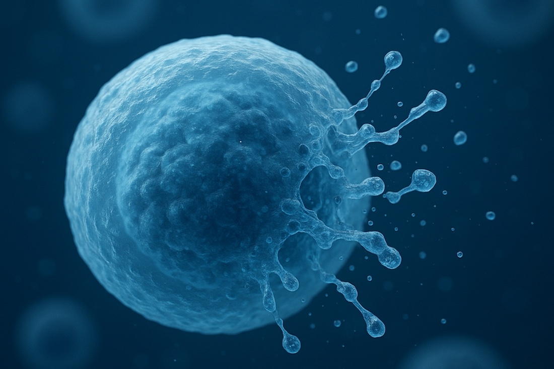
In humans and most other organisms, programmed cell death is necessary to shape tissues during development; eliminate old, damaged, infected, or unnecessary cells; or strike a balance between cell growth and death, among other functions, Dr. Najafov explained. When cells become inflamed through infection or chronic disease, they can undergo necroptosis, a form of programmed cell death in which a molecular cascade ultimately culminates in cell membrane rupture. This process releases signals that recruit immune cells to the dead cells to remove their debris and fight released bacteria or viruses.
In other forms of programmed cell death that also involve cell membrane rupture – such as apoptosis, pyroptosis, and ferroptosis – researchers have shown that a protein called NINJ1 is responsible for splitting open the cell membrane. However, NINJ1 doesn’t appear to be involved in necroptosis. Although previous studies have identified the preceding steps in the necroptosis molecular cascade, Dr. Najafov said, none had discovered a protein analogous to NINJ1 in this process.
Searching for that missing piece, Dr. Najafov and his colleagues used the gene editing tool CRISPR to eliminate individual genes in human cells that had been modified to produce an activated form of MLKL, the last known protein in the necroptosis molecular cascade. Producing this form of MLKL caused most of these cells to undergo necroptosis and burst open. The only exception was a cell clone in which CRISPR had inactivated the gene coding for a protein called SIGLEC12, which has parts that are strikingly similar to NINJ1.
When the researchers stimulated cells missing SIGLEC12 to undergo necroptosis, their cell membranes ballooned outward but didn’t rupture. Forcing cells to produce extra SIGLEC12 didn’t cause them to burst open either. A closer look showed that another protein called TMPRSS4 cuts off part of SIGLEC12, a process that seems to be key for activating it. Experiments using just this cleaved form of SIGLEC12 showed that it was sufficient to prompt cell membrane rupture.
Cells from many cancer types are less likely than healthy cells to undergo necroptosis, a factor thought to help them survive and grow. Dr. Najafov and his colleagues found that SIGLEC12 mutations, common in many cancer types, prevent this protein from being cleaved by TMPRSS4, thus stymieing SIGLEC12 function. They identified several other SIGLEC12 mutations found in the general population, which also prevent SIGLEC12 cleavage by TMPRSS4. Although the significance of these mutations isn’t known, they could affect sensitivity to infections and other inflammatory conditions, he said.
In the future, Dr. Najafov added, drugs that target SIGLEC12 or TMPRSS4 could be used to prevent necroptosis and treat conditions in which it’s a common feature.
Other UTSW researchers who contributed to this study are first author Hyunjin Noh, Ph.D., postdoctoral researcher, and Zeena Hashem, B.Sc., graduate student researcher.
This study was funded by the National Institute of General Medical Sciences (R35 GM146861) and a National Cancer Institute Cancer Center Support Grant (P30 CA142543).
About UT Southwestern Medical Center
UT Southwestern, one of the nation’s premier academic medical centers, integrates pioneering biomedical research with exceptional clinical care and education. The institution’s faculty members have received six Nobel Prizes and include 24 members of the National Academy of Sciences, 25 members of the National Academy of Medicine, and 13 Howard Hughes Medical Institute Investigators. The full-time faculty of more than 3,300 is responsible for groundbreaking medical advances and is committed to translating science-driven research quickly to new clinical treatments. UT Southwestern physicians in more than 80 specialties care for more than 143,000 hospitalized patients, attend to more than 470,000 emergency room cases, and oversee nearly 5.3 million outpatient visits a year.