UTSW study uncovers mechanisms of protein misfolding linked to neurodegenerative diseases
Understanding key protein structures could offer insights for treating and diagnosing dozens of diseases, including Alzheimer’s
DALLAS – March 27, 2023 – A team at UT Southwestern has developed a computational approach to uncover mechanisms of protein misfolding linked to neurodegenerative diseases. The study, published in Nature Communications, offers key insights that could help identify new treatments for patients.
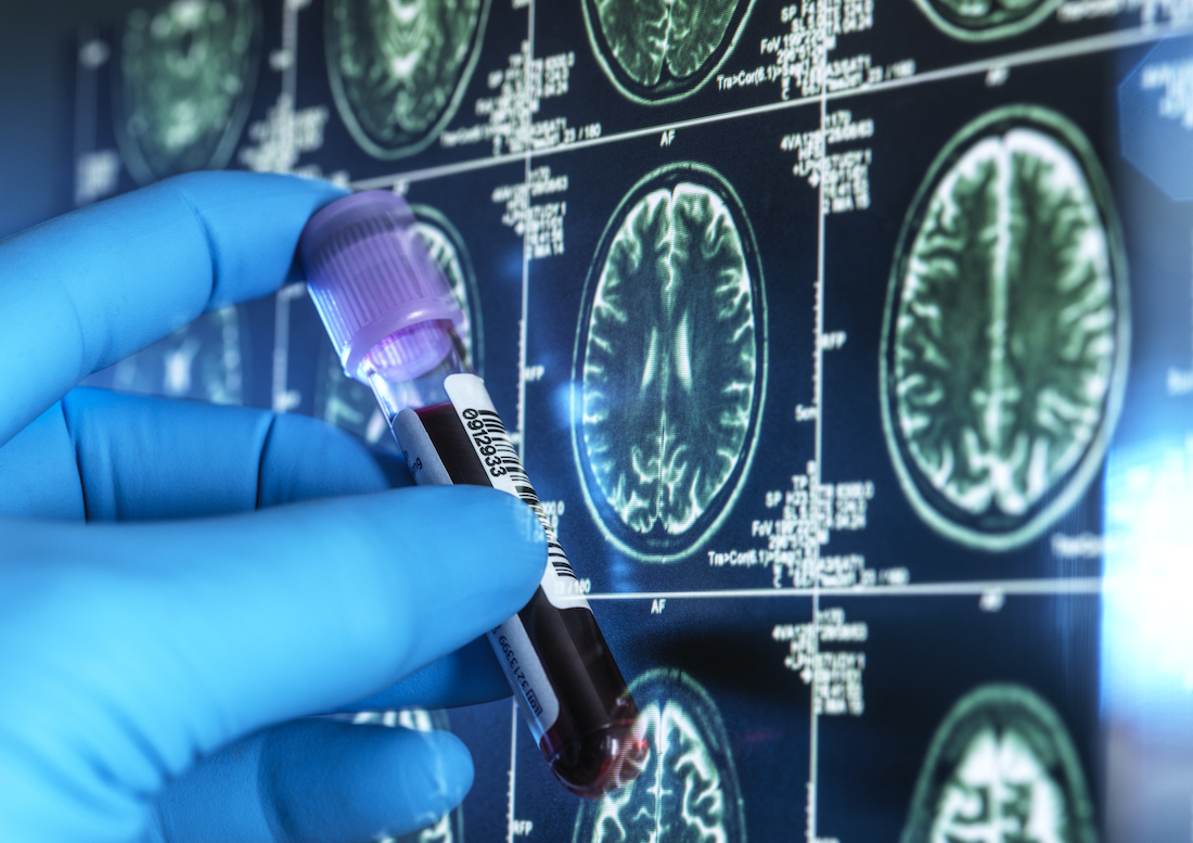
The study analyzed structures of amyloid fibrils, which are made up of proteins that are normally soluble but have assembled in a way that makes them insoluble and often dangerous. Many types of amyloids are associated with different neurodegenerative diseases.
“Understanding the mechanisms of amyloid folding will help identify novel methods to treat protein misfolding diseases,” said Lukasz Joachimiak, Ph.D., Assistant Professor of Biochemistry and in the Center for Alzheimer's and Neurodegenerative Diseases at UTSW’s Peter O’Donnell Jr. Brain Institute. Dr. Joachimiak is an Effie Marie Cain Scholar in Medical Research and the lead author of the study.
According to Dr. Joachimiak, tau proteins play key roles in healthy brain cells. However, when a tau protein misfolds in a way that exposes certain motifs that allow it to self-replicate and build on itself, it wreaks havoc in the brain. This assembly of proteins creates distinct shapes of aggregated tau proteins that are associated with neurodegenerative diseases known as “tauopathies.” The study looked at conformations from diseases classified as tauopathies, including Alzheimer’s and chronic traumatic encephalopathy, or CTE.
Using tau as a model protein, the computational approach developed by the researchers allowed them to estimate the energetics of interactions in structures of amyloid fibrils. By applying this to 27 distinct tau protein structures, they uncovered networks of interactions involved in stabilizing different amyloid fibril folds.
This information was used to classify different tauopathies based on fibril structure conformation, opening the door to design tau sequences that only adopt a single conformation. The study is believed to be the first to computationally model the effect that mutations have on fibril structure stability – an insight that will be essential in developing methods to predict these structures from the protein sequence alone.
“We are applying these methods to control folding of other amyloid-forming proteins that direct normal biological processes to begin to manipulate them,” Dr. Joachimiak said.
The study links the team’s previous work that focused on the process of misfolding in an individual tau molecule with end-stage amyloid fibril conformations observed after onset of the disease. This work was carried out in collaboration with Marc I. Diamond, Ph.D., Professor of Neurology and Neuroscience at UTSW. Dr. Diamond is the founding director of the Center for Alzheimer’s and Neurodegenerative Diseases. He also holds the Effie Marie Cain Distinguished University Chair in Alzheimer’s Research.
Other UTSW researchers who contributed to the study include Vishruth Mullapudi, Jaime Vaquer-Alicea, Vaibhav Bommareddy, Anthony R. Vega, Bryan D. Ryder, and Charles L. White III.
Dr. White holds the Nancy R. McCune Distinguished Chair in Alzheimer’s Disease Research.
This study was funded by an Effie Marie Cain Scholarship in Medical Research, The Welch Foundation (I-1928-20200401), and the Chan Zuckerberg Initiative Collaborative Science Award (2018-191983). Transmission electron microscopy was performed at the Electron Microscopy Core Facility at UTSW, which is supported by the National Institutes of Health (NIH) (1S10OD021685-01A1 and 1S10OD020103-01).
About UT Southwestern Medical Center
UT Southwestern, one of the nation’s premier academic medical centers, integrates pioneering biomedical research with exceptional clinical care and education. The institution’s faculty members have received six Nobel Prizes and include 24 members of the National Academy of Sciences, 25 members of the National Academy of Medicine, and 13 Howard Hughes Medical Institute Investigators. The full-time faculty of more than 3,300 is responsible for groundbreaking medical advances and is committed to translating science-driven research quickly to new clinical treatments. UT Southwestern physicians in more than 80 specialties care for more than 143,000 hospitalized patients, attend to more than 470,000 emergency room cases, and oversee nearly 5.3 million outpatient visits a year.