Disrupted metabolism linked to heart failure, UTSW study finds
Preclinical research reveals how overactive fat burning in heart cells destroys mitochondrial function by depleting an essential lipid
DALLAS – June 09, 2026 – When heart cells burn fat without normal metabolic controls, they can deplete a lipid needed to keep mitochondria functioning properly, according to a study by UT Southwestern Medical Center researchers. The findings, published in The Journal of Clinical Investigation, identify a mechanism linking disrupted energy metabolism to heart failure and point to potential strategies for earlier intervention.

“This study challenges a long-held assumption that maximizing fat burning is beneficial for the heart,” said senior author Jay Horton, M.D., Director of the Center for Human Nutrition and Professor of Internal Medicine and Molecular Genetics at UT Southwestern. “It demonstrates that unrestrained fatty acid oxidation (FAO) paradoxically destroys the heart’s own mitochondrial architecture through depletion of cardiolipin, an essential structural lipid.”
FAO, the process cells use to break down fats for energy, provides most of the fuel for a healthy heart. As heart failure develops, however, the heart often shifts away from fat and relies more on glucose, a pattern that has fueled debate over whether reduced FAO contributes to disease or helps defend the failing heart. The study by Dr. Horton and his colleagues suggests that under some conditions, this shift may help preserve mitochondrial function when fat metabolism becomes excessive.
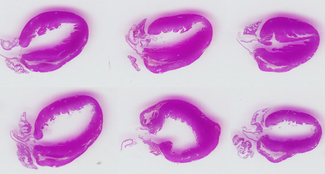
To examine what happens when the heart cannot properly regulate its fuel use, two related enzymes, acetyl-CoA carboxylase 1 and 2, were genetically removed from mouse heart muscle cells. These enzymes normally help control how many fatty acids enter mitochondria, the structures inside cells that generate energy. Without that control, the mice developed enlarged hearts and impaired blood-pumping function.
Further analysis showed how the damage unfolded. Unrestrained fat burning depleted linoleic acid, a dietary fatty acid the heart needs to maintain cardiolipin. As cardiolipin levels fell, the mitochondria’s energy-producing machinery faltered, and the mice developed dilated cardiomyopathy, a form of heart failure marked by an enlarged, weakened heart.
The study also tested whether limiting fatty acid entry into mitochondria, where FAO occurs, could change the course of disease.
“Timing matters for FAO-targeted therapy for heart failure,” said first author Chai-wan Kim, Ph.D., Assistant Professor in the Center for Human Nutrition and of Internal Medicine at UT Southwestern.

The researchers also found that drugs that inhibit CPT1, a protein that helps move fatty acids into mitochondria, prevented heart failure when given early, before cardiac dysfunction developed. However, the same approach did not improve heart function once cardiomyopathy was established.
The preclinical results suggest that therapies targeting heart metabolism may be most effective before significant cardiac dysfunction develops. They also highlight cardiolipin and related mitochondrial lipids as possible markers of risk or targets for new treatments, though the researchers said more study is needed to determine whether the same mechanisms occur in patients.
The study builds on earlier work from the Horton Lab showing that blocking acetyl-CoA carboxylase enzymes can reduce fat buildup in the liver. By studying those enzymes in the heart, the new research shows how the effects of altering fat metabolism can differ sharply by organ.
“This work suggests that the goal should not be simply to increase or decrease FAO. Instead, the heart needs metabolic flexibility that keeps FAO within a healthy range,” Dr. Horton said. “This balance is important for both energy production and mitochondrial membrane health, and it may guide future approaches for treating heart failure.”
The findings may also inform future studies of heart disease linked to obesity, Type 2 diabetes, and metabolic syndrome, conditions that can expose the heart to chronic lipid overload.
Other UTSW researchers who contributed to this study are Goncalo Dias do Vale, Ph.D., Assistant Professor in the Center for Human Nutrition and of Internal Medicine; Xiaorong Fu, Ph.D., Associate Professor in the Center for Human Nutrition and of Internal Medicine; Jeffrey McDonald, Ph.D., Professor in the Center for Human Nutrition and of Molecular Genetics; Chao Li, Ph.D., Instructor of Internal Medicine; Craig Malloy, M.D., Professor in the Advanced Imaging Research Center and of Internal Medicine and Radiology; Stanislaw Deja, Ph.D., Assistant Professor in the Center for Human Nutrition and of Biochemistry; Shawn Burgess, Ph.D., Professor in the Center for Human Nutrition and of Pharmacology; and Matthew Mitsche, Ph.D., Assistant Professor in the Center for Human Nutrition and of Internal Medicine.
Dr. Horton is a Distinguished Teaching Professor and holds the Distinguished University Chair in Human Nutrition, the Center for Human Nutrition Director’s Endowed Chair, and the Scott Grundy Director’s Chair.
This study was funded by grants from the National Institutes of Health (P01HL160487 and P30DK127984) and the American Heart Association (23SCEFIA1154964, 24IPA1272385, and 24TPA1297929).
About UT Southwestern Medical Center
UT Southwestern, one of the nation’s premier academic medical centers, integrates pioneering biomedical research with exceptional clinical care and education. The institution’s faculty members have received six Nobel Prizes and include 27 members of the National Academy of Sciences, 25 members of the National Academy of Medicine, and 13 Howard Hughes Medical Institute Investigators. The full-time faculty of nearly 3,400 is responsible for groundbreaking medical advances and is committed to translating science-driven research quickly to new clinical treatments. UT Southwestern physicians in more than 80 specialties care for more than 143,000 hospitalized patients, attend to more than 470,000 emergency room cases, and oversee nearly 5.3 million outpatient visits a year.