Electrical stimulation offers hope for treating spinal injuries
Device developed at UT Southwestern uses electrodes on skin to treat pain, spasticity, paralysis
DALLAS &ndash May 21, 2025 – A grid of electrodes placed on the backs of study participants delivered enough low-voltage electrical stimulation through the skin to change the short-term function of spinal cord neurons, a study led by UT Southwestern Medical Center researchers showed. Their findings, published in the Journal of Neural Engineering, could lead to new approaches to treat pain, spasticity, and paralysis in patients, including those with spinal cord injuries and stroke, the authors said.

“The ability to differentially and noninvasively modulate spinal circuits offers a promising alternative for patients who are unable or unwilling to undergo invasive spinal stimulation procedures,” said Yasin Dhaher, Ph.D., Professor of Physical Medicine and Rehabilitation at UT Southwestern and an Investigator in the Peter O’Donnell Jr. Brain Institute. Dr. Dhaher co-led the study with UT Southwestern graduate student Hyungtaek “Tony” Kim, M.S.
Over the past decade, advances in techniques to stimulate the spinal cord with implanted electrodes have shown enormous potential, restoring one’s ability to stand and walk even when the spinal cord is severed. These devices, which work by modifying the activity of nerve cells with electricity, hold promise for treating neurological injuries and disease. However, Dr. Dhaher and Mr. Kim said, they carry the inherent risks of invasive spinal surgery – including potential infection and injury – and require a lengthy recovery period.
Some researchers have investigated delivering electrical stimulation to the spinal cord noninvasively through electrodes placed on the skin. However, early attempts using large surface pads spread current broadly across the back, so only a diffuse field reached the spinal segments that control leg muscles, achieving minimal or no results.
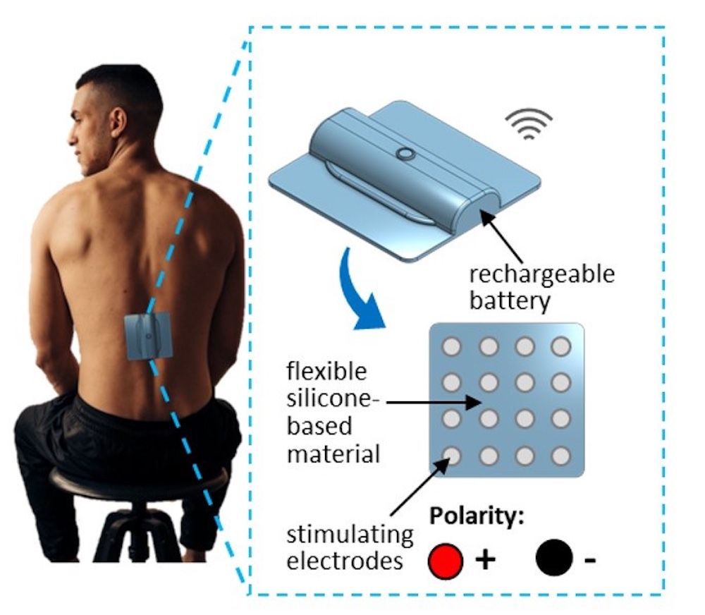
To create a “localized” field, Dr. Dhaher, Mr. Kim, and their colleagues developed an electrode grid featuring eight pairs of 1.27-centimeter anodes and cathodes arranged in a 4-by-4-inch array and attached to a biocompatible adhesive substrate. By altering which electrodes served as anodes or cathodes, the same patch could steer current either straight across (transverse layout) or diagonally across the thoracolumbar cord.
Because spinal neurons run in many directions, the team investigated field orientation that would interact most effectively with the lumbar circuits. Comparing the two layouts allowed them to see how subtle shifts in electric-field direction reshape spinal activity even with low currents.
The team recruited 17 healthy participants with an average age of about 29 to test the devices, centering the grid over the participants’ 10th and 11th thoracic vertebrae. This corresponds to the portion of the spinal cord known to control the tibialis anterior (TA), a muscle that runs along the shin and controls ankle flexion.
After checking the participants’ reflexes to confirm normal activity of the TA, the researchers delivered 40 milliamperes of electricity to the grid – about half what is needed to power the flashlight on a phone. Although this low voltage isn’t enough to stimulate neurons to cause the TA to contract, Mr. Kim explained, it changed the neurons’ excitability, or ability to fire. The researchers found that this stimulation had an inhibitory effect, lowering excitability even 30 minutes after the researchers turned off the electrode grid. This inhibition was more pronounced when the researchers positioned the grid in a diagonal orientation with corners pointing up and down the spine, rather than a transverse orientation with edges at the top and bottom.
The device was safe and easy to use, Mr. Kim said, and changing its position could allow patients to personalize its effects to treat their conditions. Although this study investigated the electric field’s inhibitory effects – which might be used to treat pain and spasticity – delivering electricity in other ways could make neurons more excitable, potentially enhancing patients’ ability to flex muscles. Being able to flex the TA could remedy foot drop, Mr. Kim added, providing a new therapy to treat this common consequence of stroke.
The researchers plan to continue investigating this device and have filed for a patent.
Other UTSW researchers who contributed to this study are Subaryani Soedirdjo, Ph.D., Senior Research Associate in Physical Medicine and Rehabilitation, and Yu-Chen Chung, Ph.D., Instructor in Physical Medicine and Rehabilitation.
This study was funded by a grant from the National Institute of Arthritis and Musculoskeletal and Skin Diseases (1R01AR069176-01A1).
About UT Southwestern Medical Center
UT Southwestern, one of the nation’s premier academic medical centers, integrates pioneering biomedical research with exceptional clinical care and education. The institution’s faculty members have received six Nobel Prizes and include 24 members of the National Academy of Sciences, 25 members of the National Academy of Medicine, and 13 Howard Hughes Medical Institute Investigators. The full-time faculty of more than 3,300 is responsible for groundbreaking medical advances and is committed to translating science-driven research quickly to new clinical treatments. UT Southwestern physicians in more than 80 specialties care for more than 143,000 hospitalized patients, attend to more than 470,000 emergency room cases, and oversee nearly 5.3 million outpatient visits a year.