Protein inhibits development of COVID-19 in live animals
Naturally occurring protein LY6E prevented mice from being sickened by SARS-CoV-2, UTSW researchers find
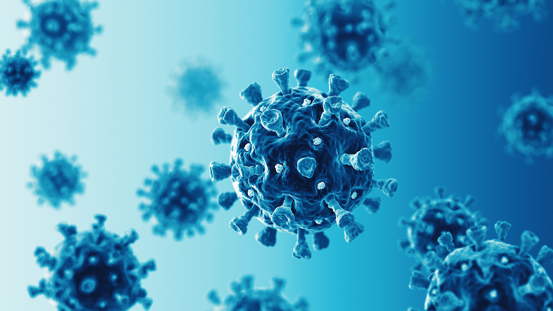
DALLAS – July 31, 2023 – A mammalian protein previously shown by UT Southwestern microbiologists to inhibit the virus that causes COVID-19 in cell culture also protected live mouse models, significantly limiting infection in the lung cells and diminishing the symptoms. The findings, published in Nature Microbiology, could lead to new strategies to treat COVID-19, which still infects thousands and kills hundreds in the U.S. every week.
“Our 2020 paper showed that LY6E could inhibit SARS-CoV-2 in cultured cells. But the proof is showing that it has the same role in living models,” said study senior author John Schoggins, Ph.D., Associate Professor of Microbiology at UT Southwestern. “We have demonstrated for the first time that the naturally occurring antiviral protein LY6E reduces COVID-19 illness in a living model.”
Human bodies use a variety of strategies to fight viral infections, including producing antiviral proteins, which are the focus of the Schoggins lab. In 2020, he and colleagues discovered that one of these proteins, LY6E, blocked infection in cell culture against a variety of coronaviruses, including one specific to mice called mouse hepatitis virus (MHV), as well as the viruses responsible for the severe acute respiratory syndrome (SARS) outbreak in 2003 and the Middle East respiratory syndrome (MERS) outbreak in 2012. But how LY6E protects against coronavirus infection and whether it could perform the same feat in COVID-19 animal models was unknown.
To answer these questions, Dr. Schoggins and colleagues used a genetic technique to generate seven live mouse models, each one engineered to turn LY6E off in specific immune cells, in all immune cells, or throughout the body. Mice missing this protein throughout their bodies or in all of their immune cells typically died of MHV infection. The mice models also became more vulnerable when the gene was turned off only in immune cells (B cells or monocytes), demonstrating the importance of LY6E in these cells for fighting coronavirus infections.
Mouse models without LY6E but infected with SARS-CoV-2 developed moderate symptoms including weight loss and inflammation and bleeding in their lungs, all signs of illness not seen in models with typical levels of LY6E.
A genetic analysis of the lungs showed that epithelial cells had significant changes in gene expression. A closer look showed that two subsets of these cells, called club cells and ciliated cells, appeared to take the brunt of infection in models without LY6E. The findings suggest the LY6E produced by these lung epithelial cell types is critical for limiting SARS-CoV-2 infection, said Dr. Schoggins, a Nancy Cain and Jeffrey A. Marcus Scholar in Medical Research, in Honor of Dr. Bill S. Vowell.
In the future, he added, researchers may be able to develop treatments for COVID-19 that depend on delivering extra genes that make LY6E or mimicking its action with a drug. In addition, mice missing LY6E could be used as more realistic models for SARS-CoV-2 infection to study drugs or vaccines. Rather than developing a fatal infection as most existing COVID-19 animal models with other genetic alterations do, or no symptoms as mice with normally functioning LY6E do, mice missing LY6E get a mild to moderate COVID-19-like illness that more closely imitates most cases of the disease in humans.
Postdoctoral researcher Katrina Mar, Ph.D., now of the Memorial Sloan Kettering Cancer Center and Institute, led the study. Other UTSW researchers who contributed were Alexandra I. Wells, Ph.D., Postdoctoral Researcher; Marley C. Caballero Van Dyke, Ph.D., Research Scientist; Jennifer L. Eitson, Senior Research Associate; Natasha W. Hanners, M.D., Assistant Professor of Pediatrics; Bret M. Evers, M.D., Ph.D., Assistant Professor of Pathology and Ophthalmology; and John M. Shelton, Lab Manager.
About UT Southwestern Medical Center
UT Southwestern, one of the nation’s premier academic medical centers, integrates pioneering biomedical research with exceptional clinical care and education. The institution’s faculty members have received six Nobel Prizes and include 24 members of the National Academy of Sciences, 25 members of the National Academy of Medicine, and 13 Howard Hughes Medical Institute Investigators. The full-time faculty of more than 3,300 is responsible for groundbreaking medical advances and is committed to translating science-driven research quickly to new clinical treatments. UT Southwestern physicians in more than 80 specialties care for more than 143,000 hospitalized patients, attend to more than 470,000 emergency room cases, and oversee nearly 5.3 million outpatient visits a year.