Radiology News
UT Southwestern Radiology Welcomes Incoming Residents
The Department of Radiology at UT Southwestern Medical Center is proud to welcome a new class of trainees following the 2026 Match Day celebration.
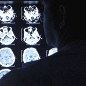
David Fetzer, M.D.: Seeing Images as Data
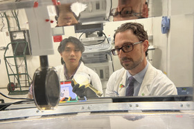
David Fetzer, M.D., likes to say that images are data. That perspective informs his work in radiology and how he approaches ultrasound at UT Southwestern.
From Chemistry to Clinical Care: Xiankai Sun, Ph.D., and Radiopharmaceutical Innovation
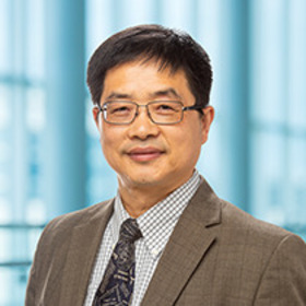
In the aftermath of China’s Cultural Revolution, as the surrounding “ridges of the Hills Everlasting” were crumbling, young students suddenly found a universe of ideas at their fingertips. Among those inspired by that moment was Xiankai Sun, Ph.D., now Director of the Cyclotron and Radiochemistry Program in the Department of Radiology at UT Southwestern.
Faculty Research Day Highlights Innovation Across Radiology
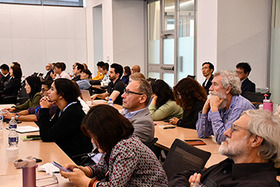
UT Southwestern’s Radiology Department hosted its annual Faculty Research Day on Wednesday, November 19, 2025, bringing together faculty for a fast-moving program highlighting scientific discover currently underway across the department.

UT Southwestern Radiology Celebrates 2025 Graduates
The UT Southwestern Department of Radiology honored 60 graduating trainees during its 2025 Graduation & Awards Celebration.
Celebrating Match Day: Welcoming Our Newest Radiology Residents
On March 18, 2022, UT Southwestern Radiology proudly celebrated Match Day—a pivotal milestone that marks the next generation of radiology talent joining our department.
Residents select new leaders for 2021
Chief Residents and Junior Chief Residents chosen by colleagues because of strong educational and administrative skills
Longtime faculty member Wally Hooser dies at 75
Dr. Hooser was a genial mentor, generous philanthropist, and space enthusiast