
Associated Cores
State of the art core facilities within the radiology department and UT Southwestern cores are essential in meeting the research objectives of the CMBQI Program.
- Cyclotron & Radiochemistry
Cyclotron & Radiochemistry
The Cyclotron and Radiochemistry Program is a UT Southwestern Medical Center-wide effort to develop the full capability of nuclear imaging, namely radioisotope-based imaging, for noninvasive assessment of physiological processes and abnormalities in animal models and in humans.
With a biomedical cyclotron and the capability to synthesize a variety of biomedical radioisotopes, this program leverages the cutting-edge technology of positron-emission tomography (PET) to enable discoveries that span multiple areas of medicine and physiology.
Visit the Cyclotron and Radiochemistry Program's website.
- Magnetic Resonance Imaging (MRI)
Magnetic Resonance Imaging (MRI)
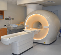
The Magnetic Resonance (MR) core is established to facilitate research and development within the Department of Radiology and collaborating departments in the field of MR imaging (MRI) and MR spectroscopy (MRS). The MR core consists of a start-of-the-art whole-body human scanner (Philips Ingenia 3T MR) and a small animal Desktop scanner (Aspect 1T MR). Philips Ingenia is a state-of-the-art MR scanner that includes dual-transmit and digital architecture for signal reception. This scanner enables the development and evaluation of new MRI/MRS techniques for improved diagnosis and understanding of the pathophysiology of disease. Aspect 1T scanner enables sequential imaging of small animals (e.g. mice and rats), without sacrificing them, for longitudinal monitoring of disease progression and/or therapy response. Being cited next to the small animal PET/CT and SPECT/CT scanners, this allows superposition of images for multimodality analysis.
Click here to explore more about the MR Core.
- Translational Molecular Imaging Core (TMIC)
Translational Molecular Imaging Core (TMIC)
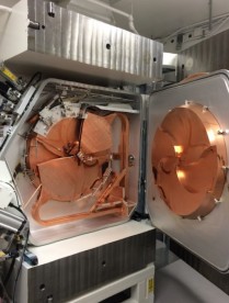
Our cyclotron and radiochemistry facility is approved for CGMP production of PET radiopharmaceuticals for human use.
- The facility includes a GE PETtrace 880 cyclotron and ancillary instruments
- Currently capable of producing 6 radioisotopes and >30 radiotracers in addition to the FDA-approved tracers
- A regulatory office in the Department of Radiology facilitates Investigational New Drug (IND) and Abbreviated New Drug Applications (ANDA) approval of radiotracers.
A GE Discovery IQ five ring PET/CT dedicated to research is available on the NE2 building, in close proximity to our cyclotron facility. This state-of-the-art digital time-of-flight (TOF) PET/CT scanner provides a 26 cm axial field of view with the highest noise equivalent count rate (NECR) of the commercially available PET/CT scanners and high NEMA sensitivity (22cps/kBq2). The five-ring system facilitates faster anatomic coverage for whole body PET/CT studies. It has both respiratory and cardiac gating capabilities. A dedicated software package (Dynamic Vue) for quantitative analysis of PET data, including 4D PET datasets, is available.
The Siemens Inveon PET/CT Multimodality System (NE3.116) is a small animal PET/CT imaging scanner built on the Siemens Inveon acquisition architecture, which fully integrates PET and CT into a common data acquisition system for automatic transition between modes and seamless coordination of CT and PET data acquisition
- Development and implementation of imaging protocols in a range of disease models (e.g., cancer, diabetes, metabolism, cardiotoxicity, neurodegenerative diseases, etc.)
- Validation of novel imaging probe development, which ranges from small organic molecules to macromolecules including proteins and nanoparticles