
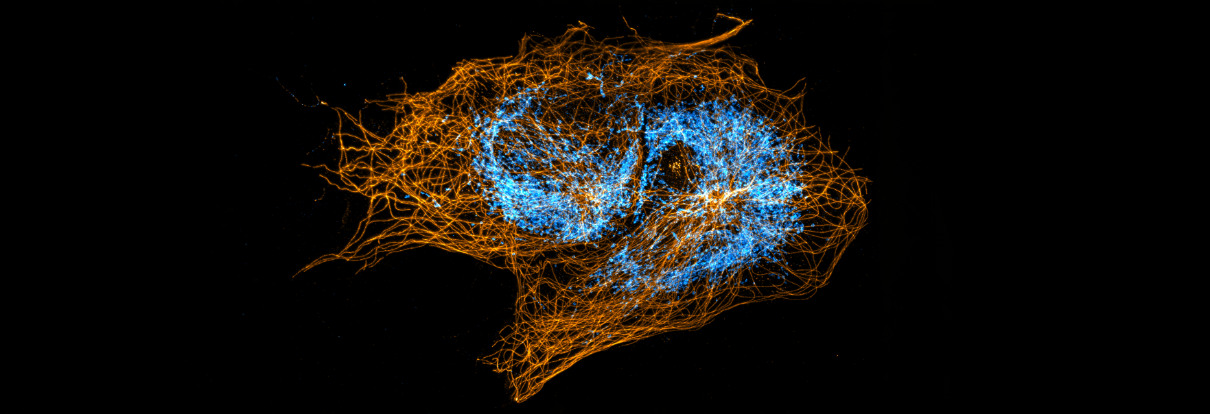
Imaging Across Biological Scales
CCIC combines tissue preparation, multiscale microscopy, expansion microscopy, and quantitative analysis to connect molecular detail with cellular interactions and whole-tissue context. Select any image to open a larger view.
Tissue architecture and biological niches
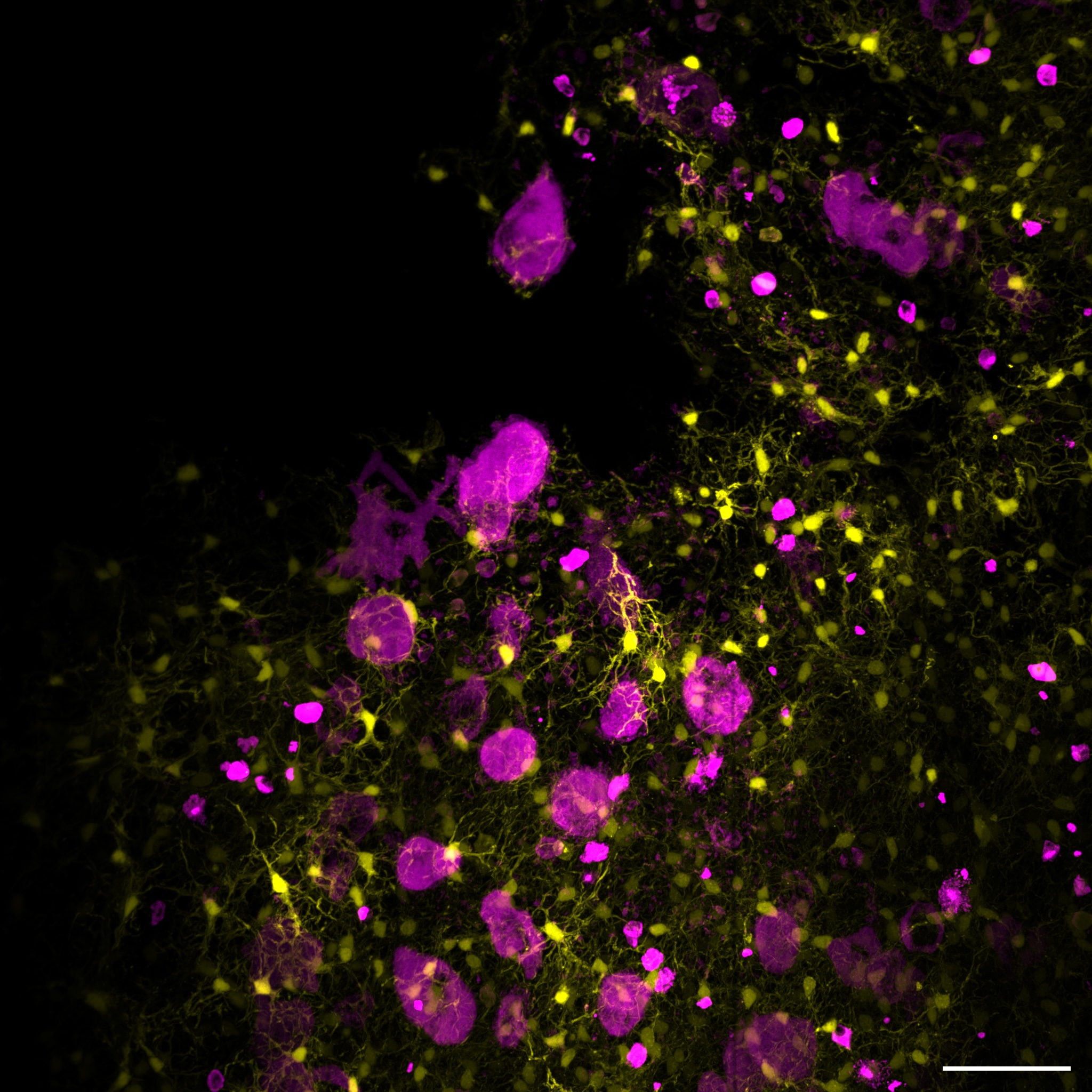
Cellular interactions in the hematopoietic stem-cell niche. Leptin-receptor-positive stromal cells interact with megakaryocytes in a spatially complex niche. Hematopoietic niche; multichannel fluorescence microscopy; 40 µm scale bar. [1]
Vasculature and whole-tissue context
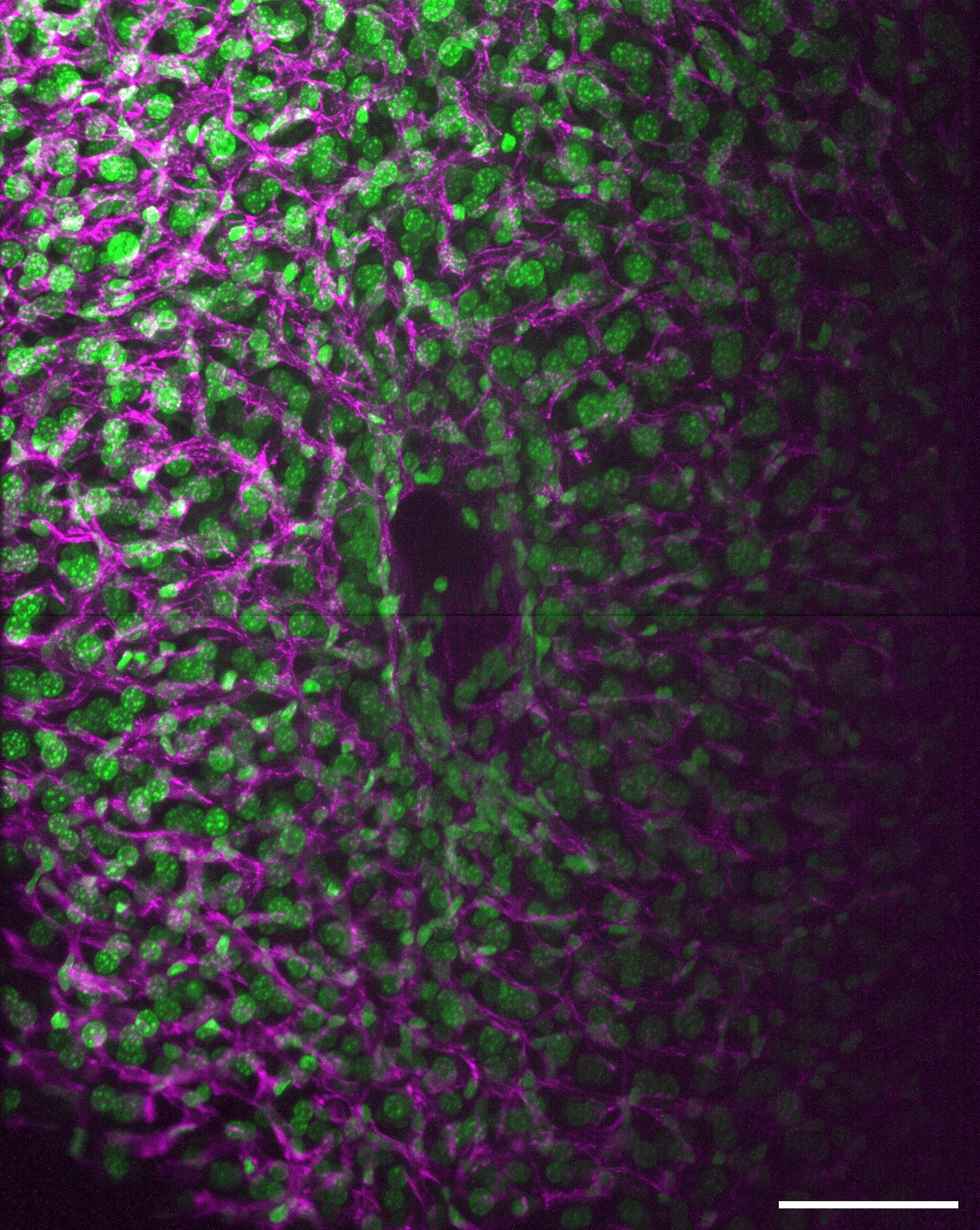
Vascular architecture in cleared mouse liver. Large-volume imaging preserves vascular organization across a thick liver specimen.
Cleared mouse liver; multichannel fluorescence imaging. [2]
Cleared mouse liver; multichannel fluorescence imaging. [2]
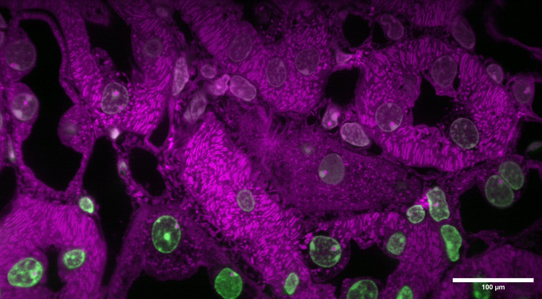
Cellular organization in mouse liver. Multichannel imaging connects cellular features with surrounding liver architecture.
Mouse liver; multichannel fluorescence microscopy. [3]
Mouse liver; multichannel fluorescence microscopy. [3]
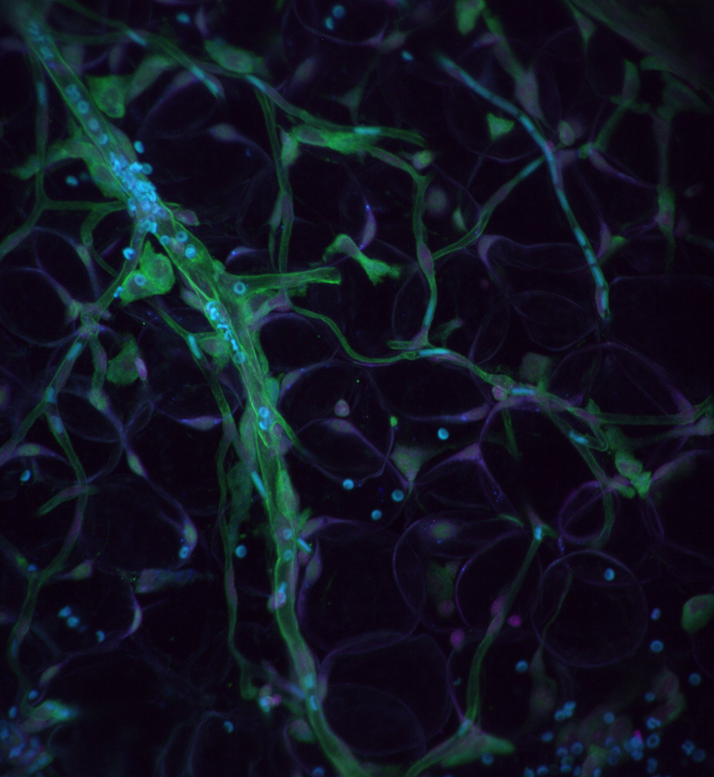
Red blood cells within a vascular network. Vascular imaging captures blood-cell flow in an adipose-rich tissue context.
Vascular network; fluorescence microscopy. [4]
Vascular network; fluorescence microscopy. [4]
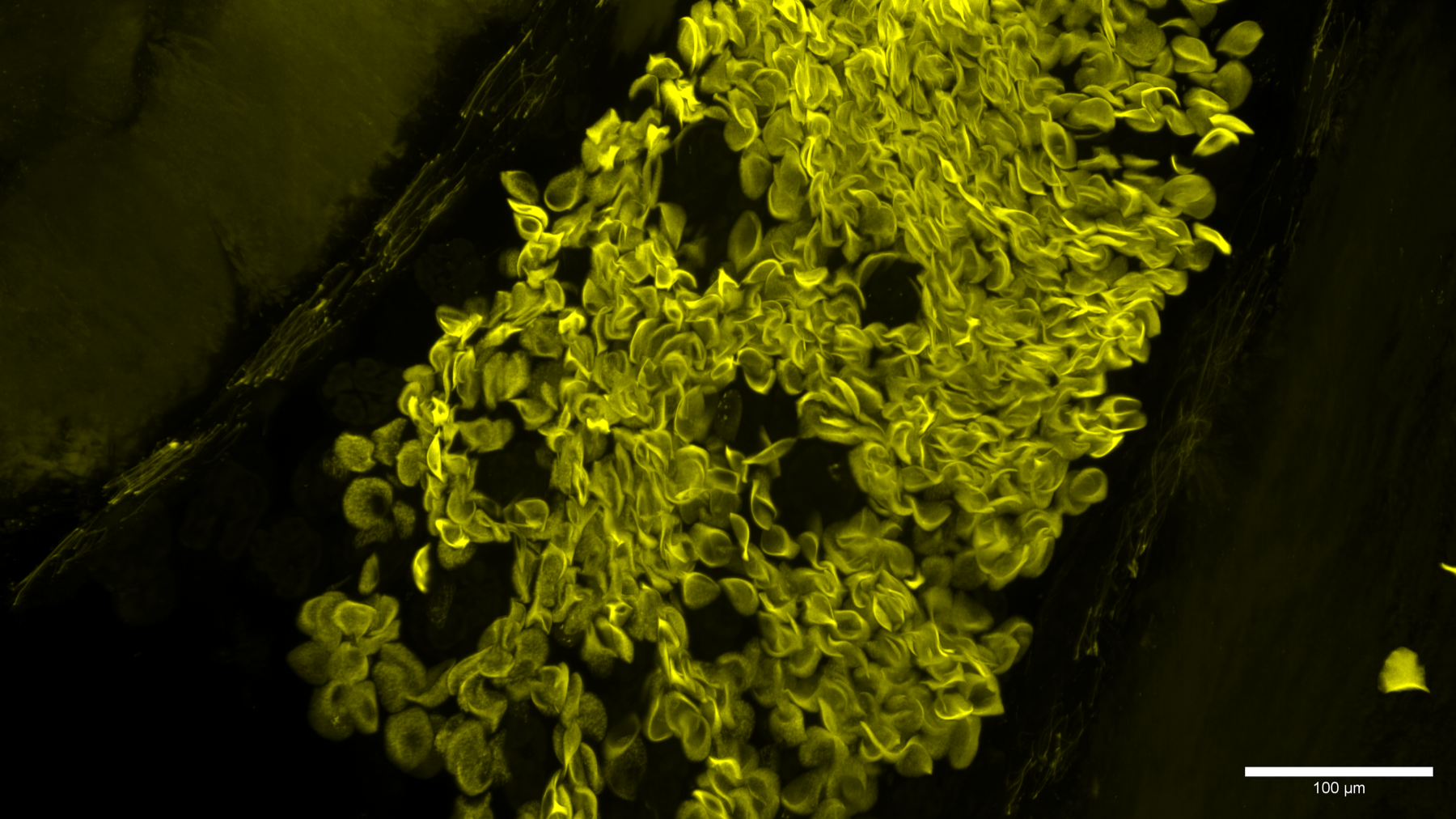
Tissue vasculature. High-resolution imaging reveals the branching organization of a tissue vascular network.
Tissue vasculature; fluorescence microscopy. [5]
Tissue vasculature; fluorescence microscopy. [5]
Cellular and organelle organization
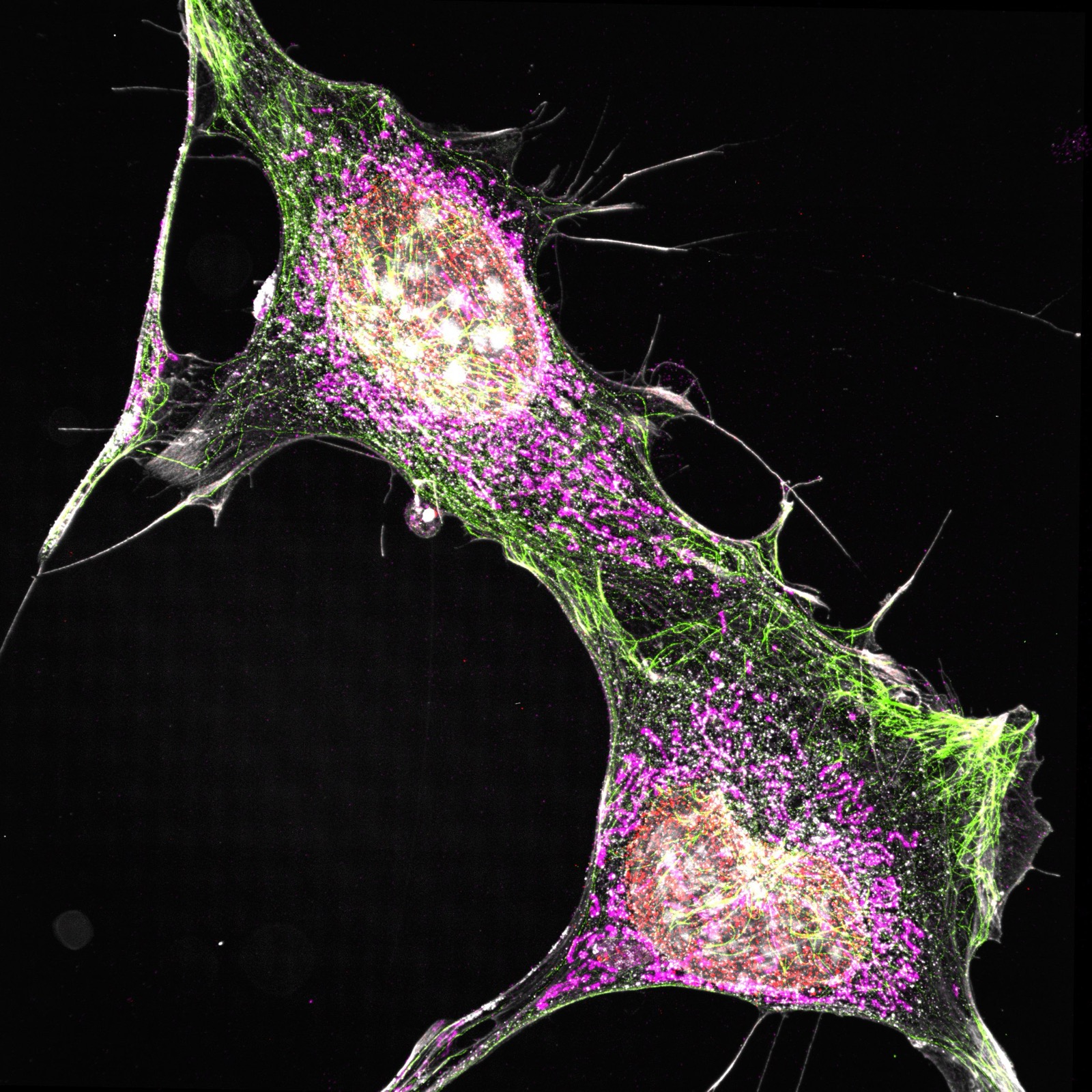
Multiplexed mouse embryonic fibroblasts. Multiplexed imaging resolves complementary cellular structures in the same field.
Mouse embryonic fibroblasts; approximately 70 × 70 × 200 nm resolution. [6]
Mouse embryonic fibroblasts; approximately 70 × 70 × 200 nm resolution. [6]
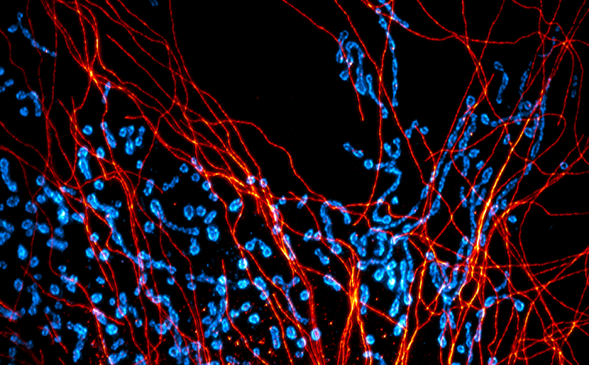
Microtubules and mitochondria in mouse embryonic fibroblasts. High-resolution imaging relates cytoskeletal organization to the mitochondrial network.
Mouse embryonic fibroblasts; approximately 70 × 70 × 200 nm resolution. [7]
Mouse embryonic fibroblasts; approximately 70 × 70 × 200 nm resolution. [7]
Expansion microscopy and molecular detail
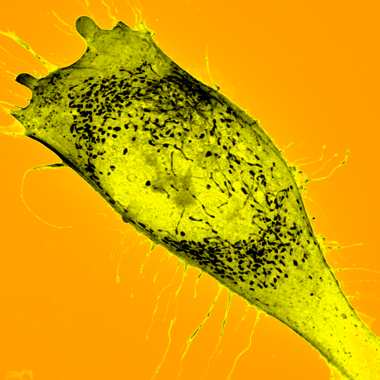
Expanded Ewing sarcoma cell. Physical expansion makes nanoscale cellular organization accessible to fluorescence imaging.
Ewing sarcoma cell; expansion microscopy. [8]
Ewing sarcoma cell; expansion microscopy. [8]
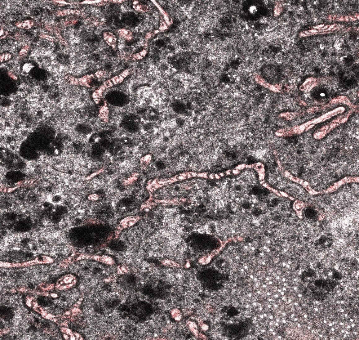
Mitochondrial cristae in a 20× expanded cell. Pan-labeling and physical expansion reveal fine mitochondrial membrane organization.
Cultured cell; 20× expansion microscopy. [9]
Cultured cell; 20× expansion microscopy. [9]
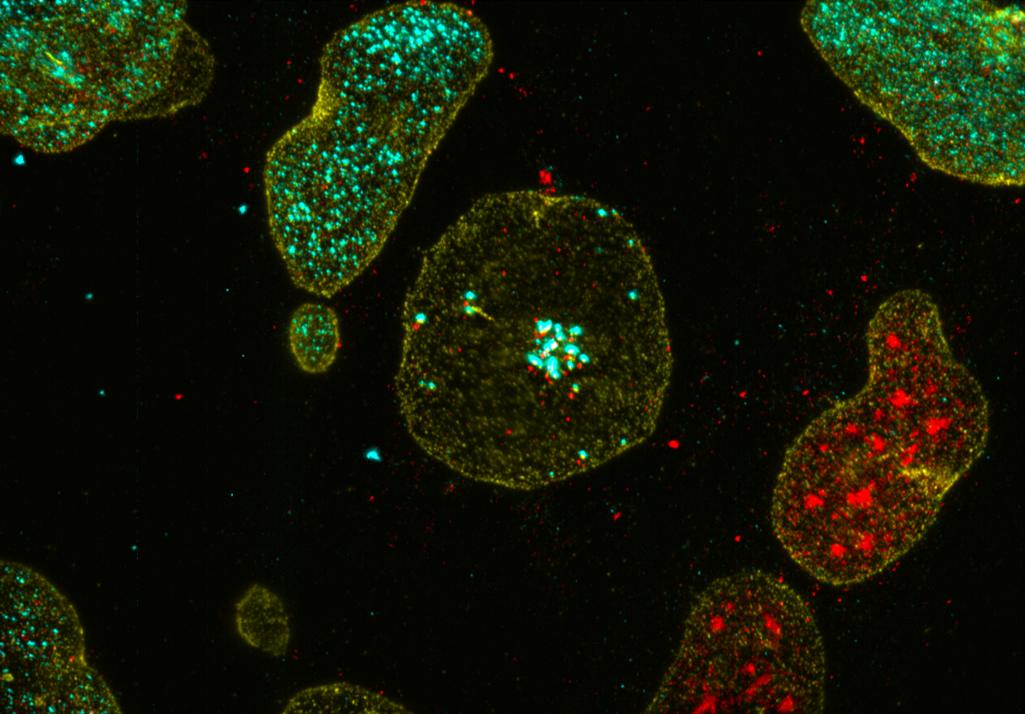
PCNA puncta in expanded cultured cells. Expansion microscopy resolves punctate PCNA organization together with RPA and the nuclear lamina.
Cultured cells; RPA, lamin, and PCNA labeling; expansion microscopy. [10]
Cultured cells; RPA, lamin, and PCNA labeling; expansion microscopy. [10]
Image credits
- In collaboration with Sean Morrison.
- In collaboration with Siyuan Zhang and Zhenyu Zhong.
- In collaboration with Hao Zhu.
- CCIC/Dean Lab.
- CCIC/Dean Lab.
- Dean Lab.
- Dean Lab.
- In collaboration with Jim Amatruda, CHLA.
- In collaboration with Joerg Bewersdorf, Yale.
- In collaboration with Peter Ly.

Supported by CPRIT. CCIC is supported in part by the Cancer Prevention and Research Institute of Texas through Core Facility Support Award RP250571.