Contact Us
T. Boone Pickens Biomedical Building
11th floor, Room 200
6001 Forest Park Road
Dallas, TX 75235
Start a Project
All new project intake goes through the UTSW iLab portal. Use iLab to request CCIC services, describe your project, and begin scheduling or consultation for sample preparation, imaging, and analysis support.
For general questions, email CCIC at tccic@utsouthwestern.edu.
What to Include in iLab
Include enough project context for CCIC staff to route the request to the right sample preparation, imaging, and analysis workflow.
- Sample type, tissue, species, fixation, clearing, expansion, or live-imaging requirements
- Biological question, markers or stains, and any antibody validation needs
- Preferred imaging service, such as cleared tissue light-sheet imaging, expansion microscopy, thick-section cyclic immunofluorescence, live spheroid imaging, or image analysis
- Expected output, such as 3D image volumes, stitched data, segmentation, visualization, or quantitative feature extraction
- Timeline, number of samples, BioHPC or data-transfer needs, and any external institution, IRB, or material-transfer considerations
Consultations and External Users
CCIC staff can help align sample preparation, microscope selection, acquisition strategy, data handling, and analysis deliverables before a project begins. Project-specific quote or rate guidance can be discussed after intake through iLab.
CCIC serves UT Southwestern investigators and cancer researchers at collaborating Texas institutions. External users should start through iLab and contact CCIC with questions about access, project fit, sample transfer, or institutional requirements.
Representative Imaging Gallery
Representative CCIC microscopy examples show the range of spatial scales supported by the core, from cell-scale structure to larger tissue contexts.
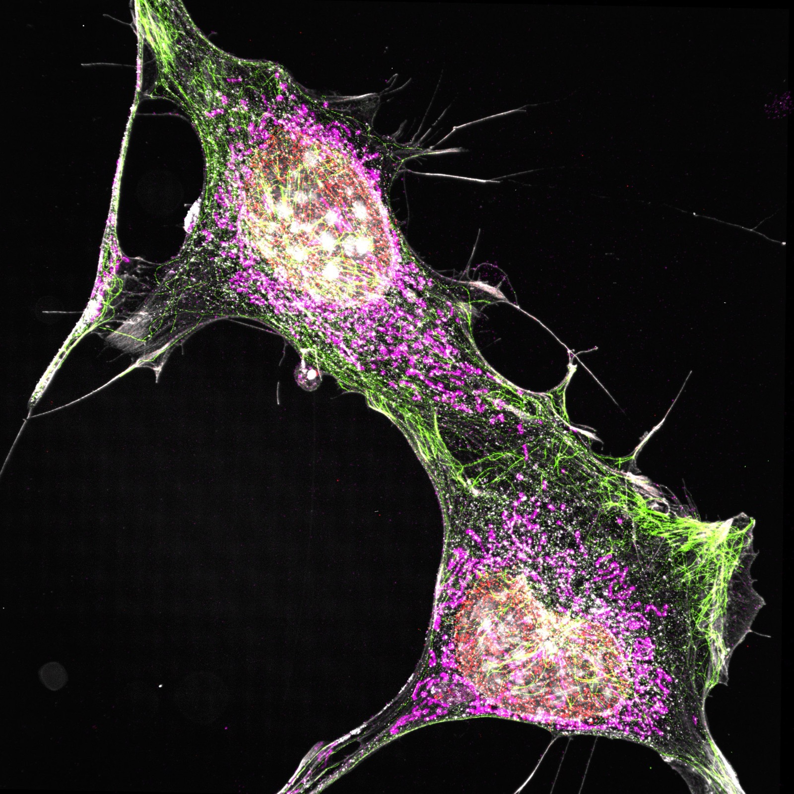
Multichannel Microscopy
Representative fluorescence imaging for cell-scale structure and spatial organization.
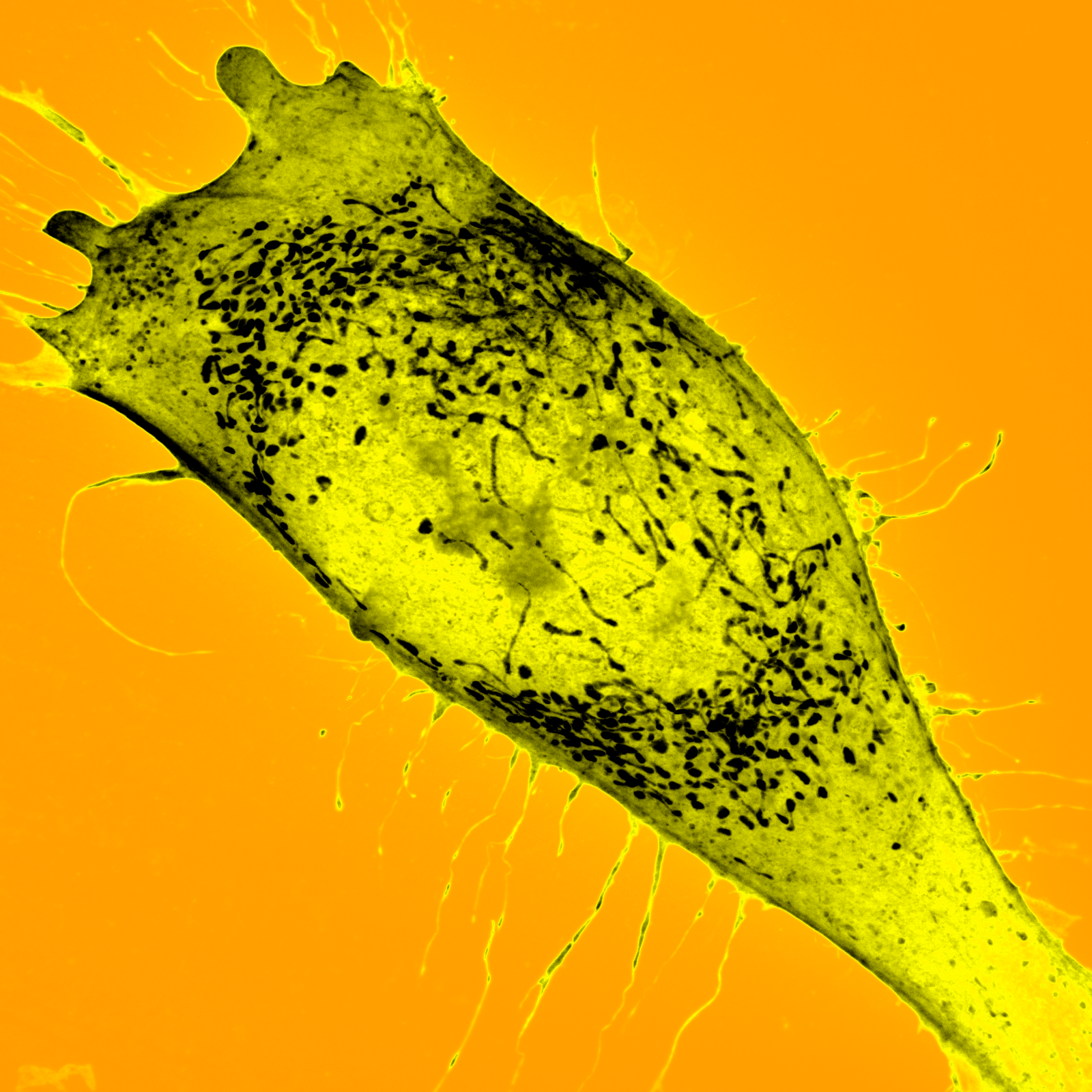
Cell-Scale Detail
Representative image highlighting subcellular and cellular morphology.

Spatial Organization
Representative fluorescence image for mapping cellular features in context.
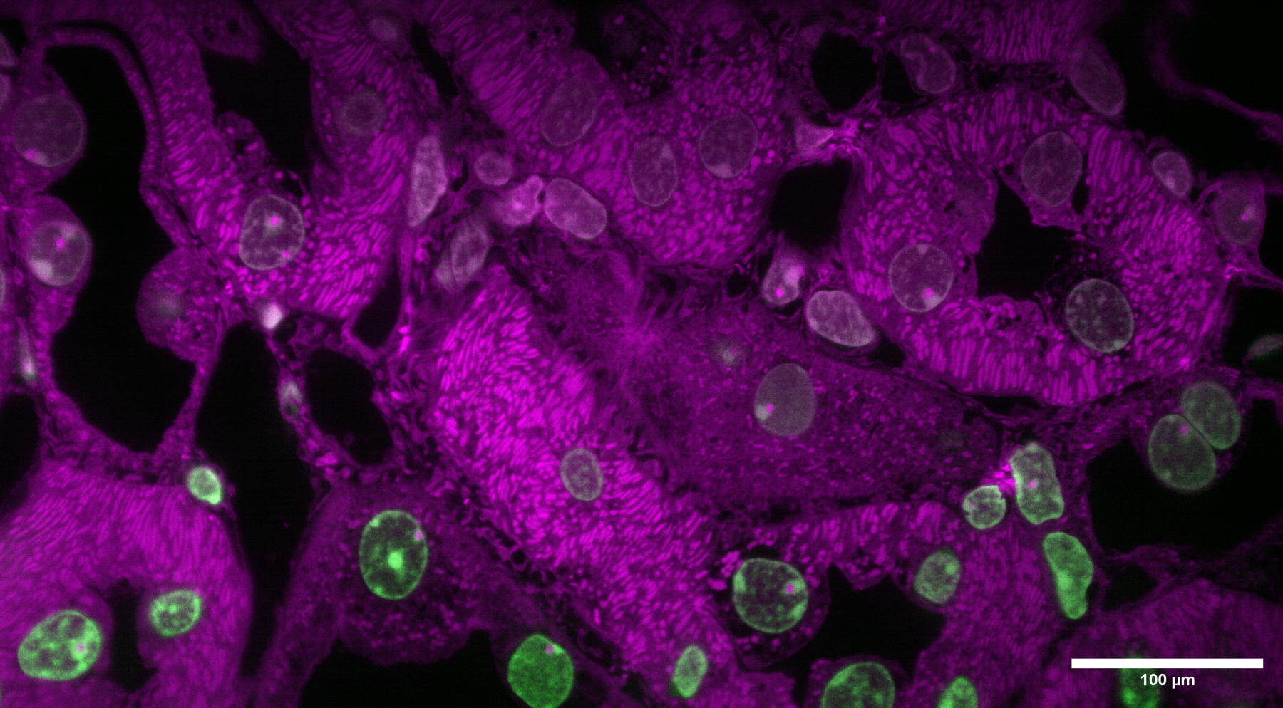
Tissue Context
Representative imaging example for evaluating structures across broader fields of view.
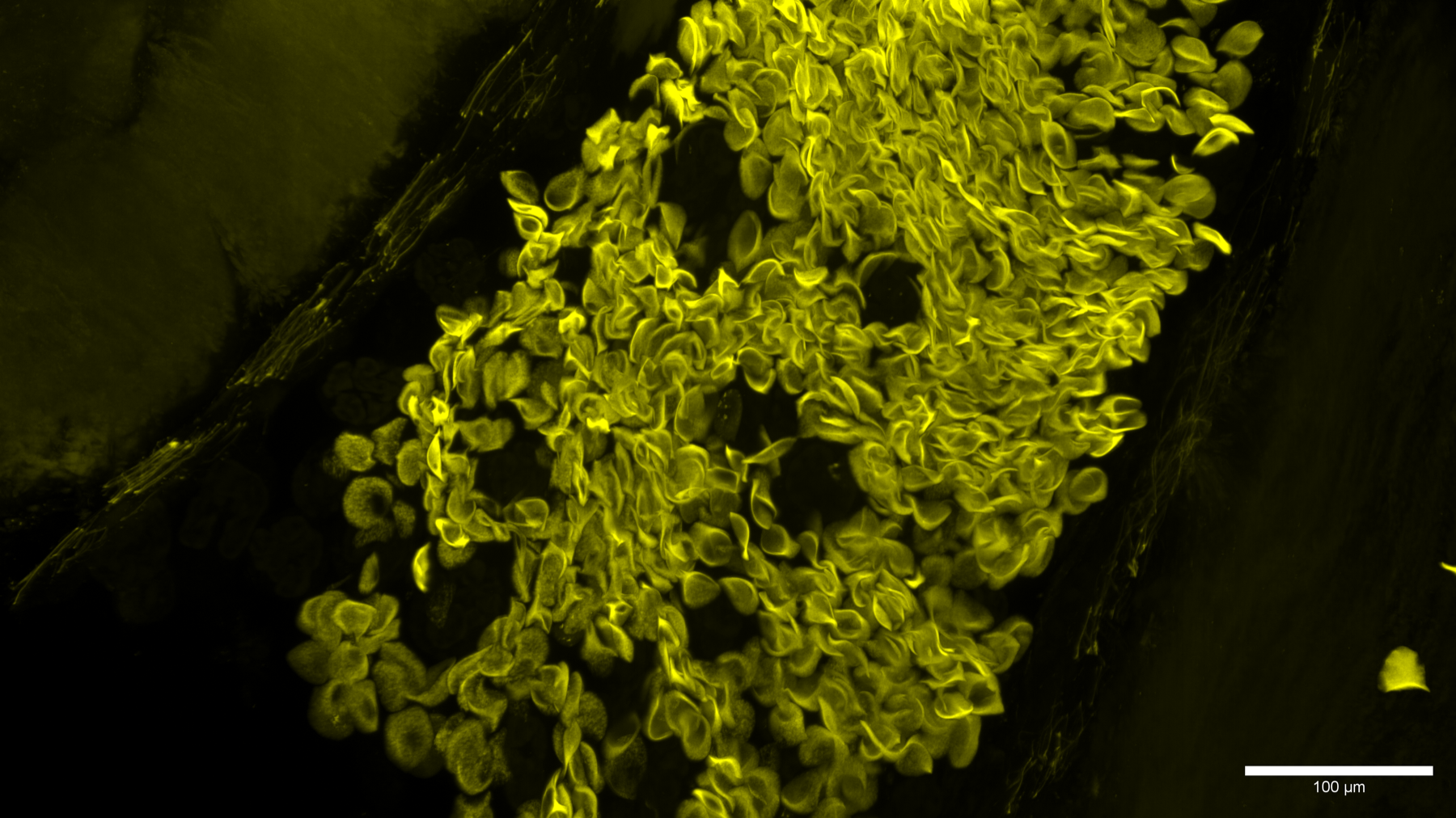
High-Resolution Imaging
Representative microscopy example with scale-bar context for detailed spatial analysis.