
Colonic/Small Intestinal LPL — Myeloid Macrophage Identification Panel
- Tissue: Colon/small intestine
- Species: Mouse
- Instrument: Cytek Aurora or BD Analyzer
- Colors: 4 (screening panel)
- Application: Myeloid/macrophage identification
Antibody Panel
| # | Target | Clone | Fluorochrome | Laser/detector | Role/cell population |
|---|---|---|---|---|---|
| 1 | Visability dye | -- | Ghost dye 710 | Red (R4) → ~710 nm | Dead cell exclusion. Ghost dye = live cells |
| 1 | CD45 | 30-F11 | BUV395 | UV 355 nm → ~395 nm | Pan-leukocyte marker. Gate CD45⁺ leukocytes from stromal cells. |
| 2 | CD64 (FcγRI) | X54-5/7.1 | StarBright Blue 700 | Blue (B9) → ~700 nm | Macrophage-specific Fc receptor. CD64⁺ distinguishes macrophages from monocytes. |
| 3 | CD11b (Mac-1) | M1/70 | FITC | Blue 488 nm → ~525 nm | Myeloid cell marker. CD64⁺ CD11b⁺ = lamina propria macrophages. |
Gating Strategy
| Step | X-axis | Y-axis | Gate | Population & Criteria |
|---|---|---|---|---|
| 1 | FSC-W | SSC-W | Scatter Gate | Exclude debris and aggregates. Intact cell cluster. Gate: ~99.9% of events. |
| 2 | SSC-A | SSC-H | Single Cells 1 | Doublet exclusion on diagonal (SSC-A ≈ SSC-H). ~90.9% of scatter gate. |
| 3 | Ghost 710 (AF700) | CD45 (BUV395) | Viable CD45+ | Ghost Dye⁻ CD45⁺ = live leukocytes. Upper-left quadrant. ~45% of single cells (78,563 events). |
| 4 | CD64 (SBB700) | CD11b (FITC) | Macrophages | CD64⁺ CD11b⁺ double-positive = lamina propria macrophages. ~19.1% of viable CD45⁺. |
Gating Hierarchy
- Scatter (FSC-W/SSC-W)
- Singlets (SSC-A/SSC-H)
- Viable CD45⁺ (Ghost 710⁻ CD45⁺)
- Macrophages (CD64⁺ CD11b⁺)
See the graphic below for representative gating plots.
Sequential Gating Strategy — LPL Macrophage Identification
Small Intestinal Lamina Propria: Representative density plots show the four-step myeloid macrophage gating workflow.
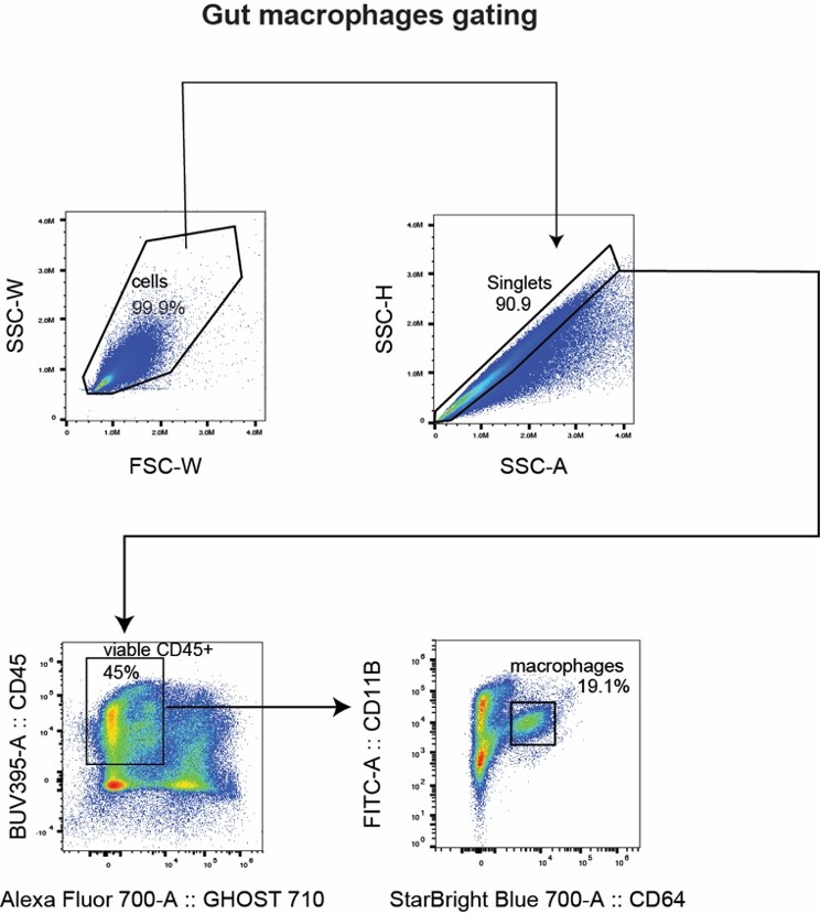
Figure: Representative flow cytometry density plots from small intestinal lamina propria. Each plot shows the gate applied at that step and the percentage of the parent population captured. Gate 4 (CD64⁺ CD11b⁺) identifies tissue macrophages and represents approximately 19% of viable CD45⁺ leukocytes.
Intestinal Lamina Propria (LPL) Isolation Protocol
Tissue-specific note:
- Small intestinal LPL: HBSS + 3% FCS + 20 mM EDTA for the epithelial stripping step. Remove Peyer's patches before digestion.
- Colonic LPL: substitute 10 mM EDTA + 1 mM DTT — DTT disrupts the colonic mucus layer for better LP cell access.
- Both protocols use the same digestion and Percoll gradient steps below.
Materials
- HBSS, 37°C, filter-sterile (no phenol red, no Ca/Mg; + NaHCO₃ 4.17 mM) + 3% FCS + 20 mM EDTA
- Colonic LPL variation: substitute 20 mM EDTA + 1 mM DTT (DTT loosens mucus)
- Digestion solution: RPMI +4.17 mM NaHCO₃ +5% FCS +Collagenase IV (125 µL/50 mL) +DNase I 0.1 mg/mL (250 µL/50 mL) +Dispase II (250 µL/50 mL)
- 100 µm and 70 µm cell strainers
- 40% and 80% Percoll (room temperature)
- Gavage needle and 10 mL syringe
- Sterile 50 mL and 15 mL conical tubes
- Sterile bottles and Erlenmeyer flasks
- Steritop 0.22 µm filters
- Large volume ice-cold PBS
Procedure
- Harvest intestinal tissue; place in ice-cold PBS +3% FBS in a Petri dish on ice.
- Flush the lumen with 10 mL ice-cold PBS via gavage needle to remove luminal contents.
- Remove Peyer's patches (small intestine). Cut intestine open lengthwise with scissors.
- Cut into ~1-inch pieces. Transfer to 50 mL conical with 10 mL ice-cold 3% PBS.
- Wash tissue 2× with 10 mL ice-cold 3% PBS: vortex 5 min (~10–12 inversions), pour off supernatant, repeat.
- Epithelial stripping: add 10 mL HBSS + 3% FCS + EDTA (see tissue note above). Rotate at 250 RPM, 37°C, 15 min. IEC collection: if collecting intraepithelial cells (IECs) for flow, incubate on ice with gentle vortexing every 5 min instead.
- Vortex 2 min. Pour off supernatant (contains IELs). Repeat step 6 once more.
- Enzymatic digestion: add tissue pieces to 10 mL RPMI digestion solution (Collagenase IV +DNase I +Dispase II). Incubate 37°C, 250 RPM, 45 min. Tip: do not over-digest; 45 min is the maximum. CD64 surface expression can be reduced by excess collagenase.
- Vortex 2 min. Pass supernatant through 100 µm → 70 µm cell strainers into a 50 mL conical.
- Second digestion of remaining tissue: 30 min, 37°C, 250 RPM. Pool supernatants from both digestions.
- Centrifuge pooled supernatant to pellet cells. Discard supernatant.
- Percoll gradient: resuspend pellet in 10 mL room-temperature 40% Percoll. Carefully underlay with 3.5 mL 80% Percoll in a 15 mL conical.
- Centrifuge: 800 × g, 20 min, no brake, room temperature.
- Collect buffy coat from the 40%/80% interface. Wash 1–2× with ice-cold media.
- Count cells. Proceed to antibody staining per the UTSW General Flow Cytometry Staining Protocol.
Tips for optimal macrophage recovery
- Macrophages sediment below the interface — also collect just above the 80% Percoll layer when transferring.
- Collagenase IV lot variation affects digestion efficiency and CD64 surface expression. Titrate each new lot.
- If CD64 signal is weak: reduce Collagenase IV concentration or add a brief Fc block (anti-CD64 clone 93) post-isolation.
- Add CountBright or similar counting beads before acquisition for absolute cell counts.
- For sorting macrophages: substitute the Sorting Staining & Sample Prep protocol and skip fixation.