Whole Brain Microscopy Equipment
The WBMF offers access to a unique set of high-throughput microscopes and histology resources, ideal for collecting high-content neuroscience data, for UT Southwestern and external researchers.
Please remember to acknowledge your usage of our facility resources in your publications using our established Research Resource Identifier code anywhere in your methods or acknowledgements: RRID: SCR_017949. This is critical for us to maintain support for the core.
Jump to: Light Sheet Microscopy | Serial Two-Photon Tomography | Whole Slide Imaging | Small Microscopes & Sectioning Equipment | Computer Workstations
Light Sheet Microscopy
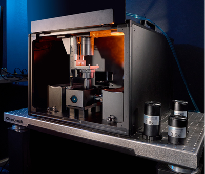
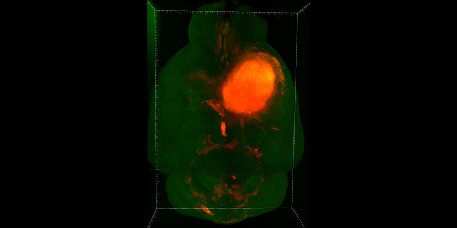
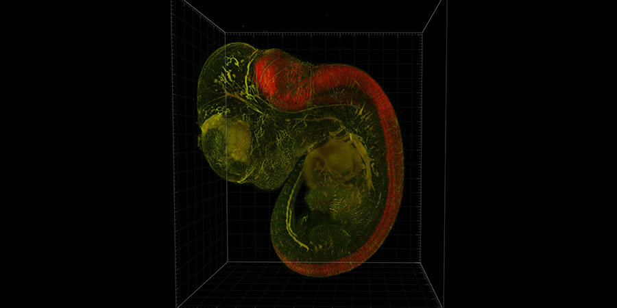
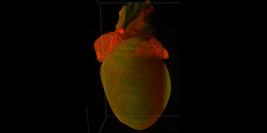
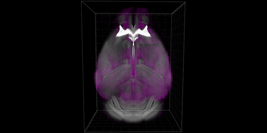
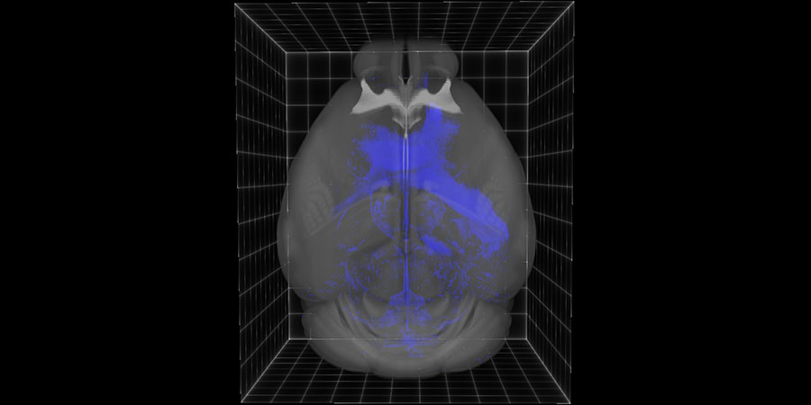
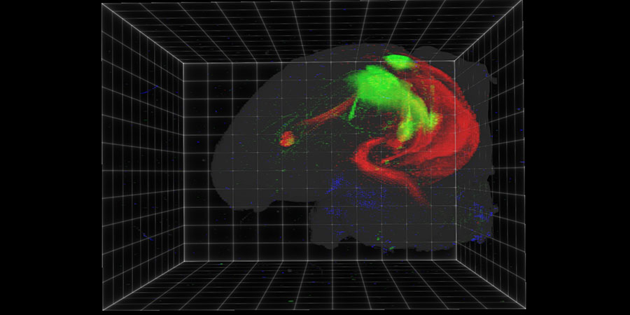
This Lightsheet microscope is designed for extremely high-speed, high-resolution volumetric imaging of optically cleared whole rodent brains or other tissues. Equipped with 1.6X, 3.6X, and 9X objectives to meet a range of imaging applications at 0.71-4um resolution. Uses patented axial sweeping technology, which enables acquisition of true isotropic 3D images and undistorted orthogonal viewing of signals of interest in any plane. The system offers focus compensation and tunable imaging parameters as well as intuitive acquisition software with real-time tile correction.
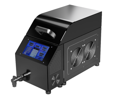
The LifeCanvas SmartBatch companion system is used for active, electrophoretic immunolabeling of whole rodent brains or other tissue samples for subsequent clearing and lightsheet imaging. The system can also be used to perform active delipidation of difficult-to-clear organs such as bone, liver, or tumor.
Serial Two-Photon Tomography
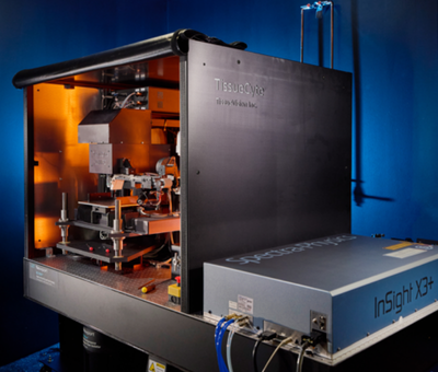
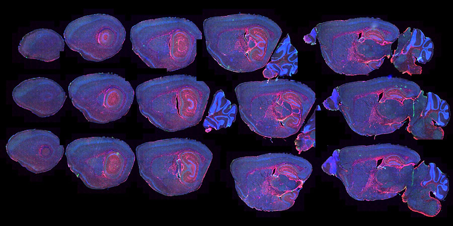
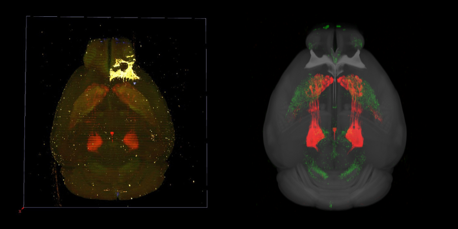
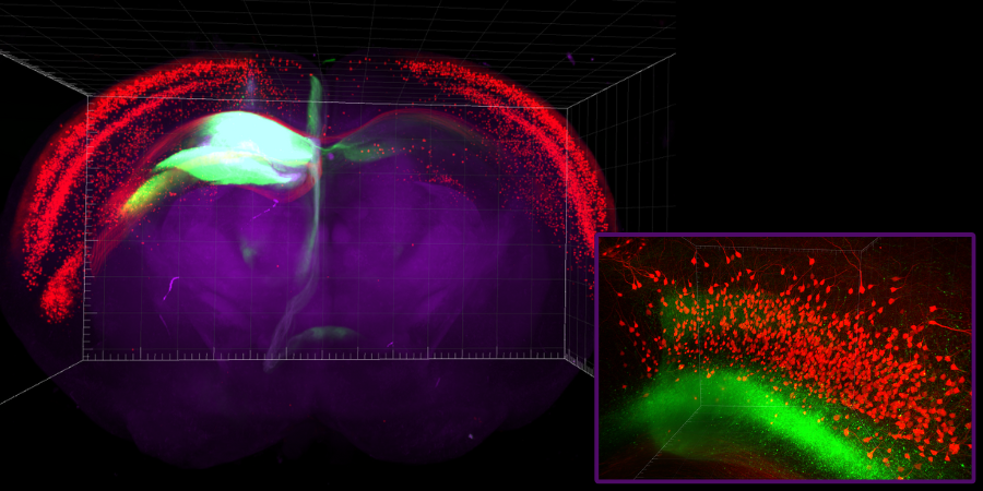
We offer a TissueVision Marinus MP serial two-photon tomography (STPT) instrument, featuring an integrated precision vibratome and multiphoton microscope, for automated sectioning and whole-brain imaging (~20 hours per mouse brain).
The system supports imaging of fluorescently labeled mouse brains, including transgenic models (e.g., GFP, tdTomato), tracer-injected brains, and chemically labeled samples for vasculature or amyloid plaques. Incorporates the SpectraPhysics Insight X3 dual line laser operating from 680–1300nm to provide greater flexibility in the choice of fluorophore and multiplexing capability. The Marinus MP also includes the robotic “section capture” device, enabling the automated collection of imaged sections for subsequent correlative staining. Perfusion-fixed brains are submitted for imaging by facility staff.
Resolutions
Typical resolutions are ~1 μm (x–y) and 25 μm (z).
Emission Bands
Up to four emission bands (blue, green, red, and far-red) are detected by independent PMTs.
Image Analysis
Our automated image analysis pipeline includes supervised machine learning for pixel classification, registration to the Allen Brain Atlas (CCF v3.0), and quantification of fluorescent signals across all brain regions. These tools enable high-throughput, standardized analysis of large experimental datasets, supported by our two full-time computational scientists.
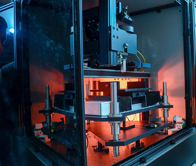
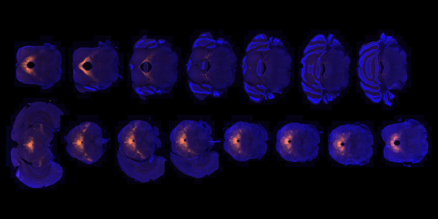
We offer a TissueVision TissueCyte 1000 serial two-photon tomography (STPT) instrument, featuring an integrated precision vibratome and multiphoton microscope, for automated sectioning and whole-brain imaging (~20 hours per mouse brain).
The system supports imaging of fluorescently labeled mouse brains, including transgenic models (e.g., GFP, tdTomato), tracer-injected brains, and chemically labeled samples for vasculature or amyloid plaques. Incorporates the SpectraPhysics MaiTai tunable laser operating from 690-1040nm and integrated laser alignment modules. Perfusion-fixed brains are submitted for imaging by facility staff.
Resolutions
Typical resolutions are ~1 μm (x–y) and 25 μm (z).
Emission Bands
Up to three emission bands (blue, green, and red) are detected by independent PMTs.
Image Analysis
Our automated image analysis pipeline includes supervised machine learning for pixel classification, registration to the Allen Brain Atlas (CCF v3.0), and quantification of fluorescent signals across all brain regions. These tools enable high-throughput, standardized analysis of large experimental datasets, supported by our two full-time computational scientists.
Whole Slide Imaging
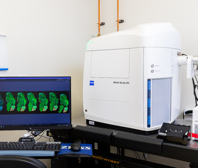
This high-speed digital slide scanner converts glass slides into high-resolution images in both brightfield and fluorescence modes. The system can automatically scan up to 100 standard-size or 50 large-format (2in by 3in) slides, using advanced cameras, precision optics, and LED light sources to deliver exceptional image quality while minimizing photobleaching.
Magnification options: scanned at 10x, 20x, or 40x resolution
Fluorescent Channels
Four independent fluorescent signals per slide: blue, green, red, and far-red.
Images are viewable in the free ZenLite software. After training, users may schedule scanning sessions or request WBMF staff assistance for an additional fee.
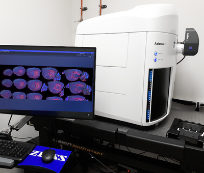
This high-speed digital slide scanner converts glass slides into high-resolution images in both brightfield and fluorescence modes. Each system can automatically scan up to 100 standard-size or 50 large-format (2in by 3in) slides, using advanced cameras, precision optics, and LED light sources to deliver exceptional image quality while minimizing photobleaching.
Magnification options: 10x, 20x, or 40x
Fluorescent Channels
Five independent fluorescent signals per slide: blue, green, red, far-red, and near-infrared.
Images are viewable in the free ZenLite software. After training, users may schedule scanning sessions or request WBMF staff assistance for an additional fee.
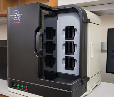
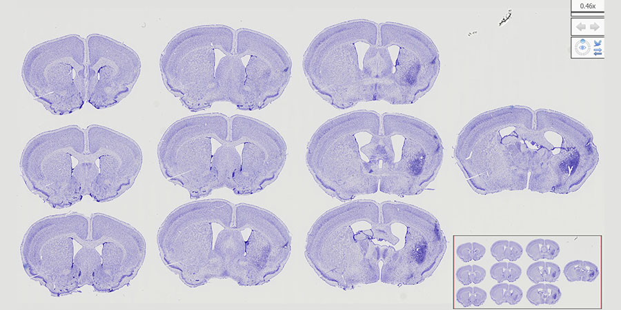
We have two NanoZoomer S60 high-speed digital slide scanners that convert glass slides into high-resolution images in about 60 seconds for a 15 × 15 mm brightfield sample at 20x. It can automatically process up to 60 standard slides or 30 large-format (2 × 3 in.) slides with excellent image quality at 20x or 40x resolution.
The Z-stack function enables multilayer scanning for viewing multiple focus planes, and images can be explored in the free NDPView2 software. After training, users may schedule scanning sessions. Alternatively, users can request full-service scanning by WBMF staff for an additional fee.
Small Microscopes and Sectioning Equipment
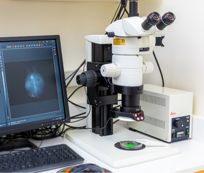
A fluorescence stereomicroscope with a digital monochrome camera. Used for imaging of whole organs and large, unmounted pieces of tissue. Optimized for fluorescent imaging using GFP or DsRed filtersets, but monochrome brightfield images can be collected as well.
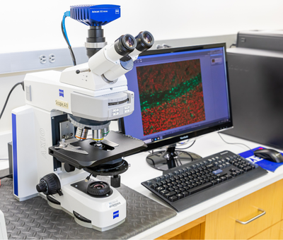
An upright epifluorescence microscope with a digital monochrome camera. Used for basic imaging of slide-mounted sections in either brightfield or fluorescence mode. The system is non-motorized. 2.5X, 10X, 20X, 40X, and 63X dry objectives are currently installed. Blue, green, red, and far-red channel fluorescence can be imaged.
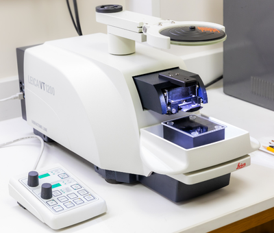
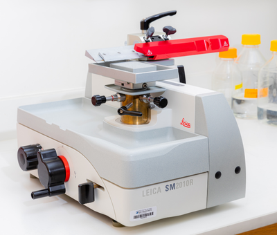
The Leica VT1200 semiautomatic vibrating blade microtome is designed for preparing thick sections of fixed, room temperature specimens (10s or 100s of microns). Can be used to optimize vibratome sectioning conditions for non-brain organs before TissueCyte serial two-photon imaging.
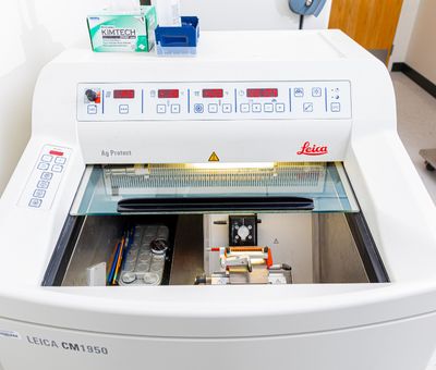
For preparing thin sections (
It can be used to prepare frozen serial sections (e.g., for rodent brain) using a dry ice tray or to cut sections of paraffin blocks.
Computer Workstations
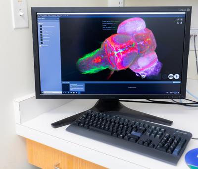
Custom-built workstation computer equipped with dual Intel Xeon Gold 2.1Ghz processors, dual Nvidia RTX8000 48GB graphics cards, 1TB RAM, and 96TB of local storage. The system is running Oxford Imaris software for 3D rendering, video creation, and analysis of high-resolution, large-volume images acquired via lightsheet microscopy. The system can also support the use of QuPath, an open source platform for AI-enabled quantification of whole slide digital images (brightfield or fluorescence).
Available Software: Oxford Imaris, QuPath, Syglass (under development)
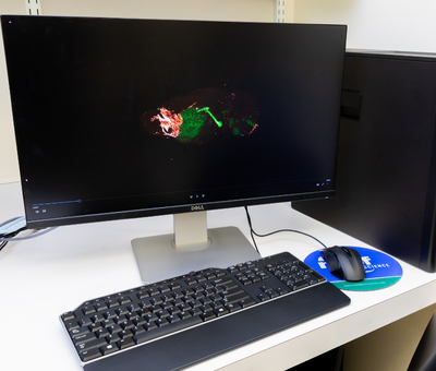
The Microbrightfield workstation runs MBF Bioscience’s Stereo Investigator and Neurolucida 360 software packages for offline stereological analysis, neuron tracing, and 3D rendering of large, whole-brain datasets. This computer can also be utilized to view and process large image files generated on the slide scanners in our facility. ImageJ/FIJI, Zeiss Zen Lite, Hamamatsu NDPView2, and Aperio Imagescope are also available for use on this computer.
Available Software: Microbrightfield Bioscience StereoInvestigator, Microbrightfield Bioscience Neurolucida 360, ImageJ/FIJI, Zeiss Zen Lite, Hamamatsu NDPView2, Leica Aperio Imagescope.
Data Images