Magnetoencephalography Core
About Us
The Magnetoencephalography (MEG) Center of Excellence features a state-of-the-art MEGIN Triux-Neo MEG system. The system is used to noninvasively and continuously measure brain activity and offers excellent temporal (
Our mission is to provide comprehensive clinical epilepsy and pre-surgical mapping MEG services to the Dallas Fort Worth community and regional patient populations, provide advanced research MEG capabilities to local universities and medical centers, and educate and train the next generation of physicians and researchers in current and emerging MEG technologies.
- Faculty & Staff
Faculty & Staff
Professor
Director, Magnetoencephalography Program
Research Lab
Technical Director, Magnetoencephalography Program
Assistant Professor
Chief of MEG Clinical Neurophysiology & Epilepsy, Magnetoencephalography Program
Assistant Professor
Amy Proskovec, Ph.D.
Assistant Professor

Adriana Ohm
MEG Technologist

Anna Manning-Mortimer
MEG Technician
- Policies & Procedures
Policies & Procedures
View our applicable policies and procedures below (requires RADpoint access):
- Resources
Resources
MEGIN Triux Neo
-
The system has 306 SQUID sensors (102 magnetometers and 204 planar gradiometers) with a dynamic range of +/- 20nT and high tolerance for magnetic interference.
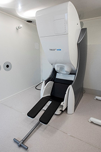
- The system has an internal closed loop helium recycler, limiting helium consumption and downtime for refills to virtually zero.
- The MEG scanner is housed in a Premium 3-layer Vaccuumshmelze magnetically shielded room with the highest shielding factors commercially available.
Stimulus Devices
All of the devices listed below are available for researchers to use during MEG scans at no additional cost. Each stimulus or response is time locked and recorded with the MEG data. E-Prime 3 is the preferred software for stimulus delivery.
- Up to 12 EKG/EOG/EMG channels
- Digitimer current stimulator
- Panasonic PT-DS12K DLP Projector
- Optical light sensor
- Right and left-hand five-finger response boxes
- Optical microphone
- SPO2 sensor
- Pressure response sensor
- Three tri-axial accelerometers
- Pneumatic headphones
EEG caps and electrodes are available for a small fee to cover the cost of disposables and technologist time (if needed).
- 64-channel EEG cap
- Loose EEG electrodes for 10-10 or 10-20 placement
- Certified EEG technologists are available to assist with placement.
Facilities
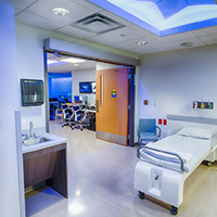
- The MEG suite has an attached "prep-room" with a hospital bed, medical gases, sink, fridge for sample storage, and supplies needed for EEG application.
- Two clinical workstations/computers are available. One in the prep-room and one in the MEG suite.
-
- Rates
Rates
Please see iLab for current rates and scheduling availability.