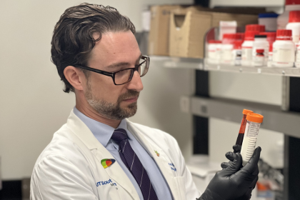
David Fetzer, M.D.: Seeing Images as Data
David Fetzer, M.D., likes to say that images are data. That perspective informs his work in radiology and how he approaches ultrasound at UT Southwestern.
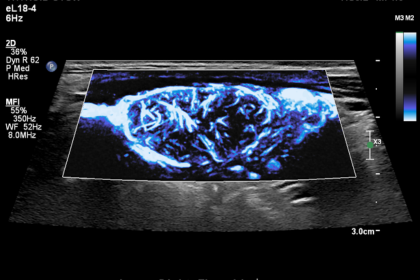
“One of the most exciting things about my work is that we are applying novel and emerging US-based techniques in a clinical setting, on clinical patients, in a variety of organs and using a variety of techniques,” Fetzer says. “From elastography, characterization of tissue attenuation and backscatter, or contrast-enhanced ultrasound qualitative and quantitative assessment, there are a wide range of US-based tools available now, with new techniques and novel applications emerging every year.”
Fetzer’s interest in ultrasound began during his training. As an undergraduate in Imaging Science, he worked on ultrasound-related research as part of his senior thesis. Later, during an Abdominal Imaging fellowship in a program strong in abdominal imaging and particularly strong in ultrasound, he built on that experience. When he first arrived at UT Southwestern, however, his attention was elsewhere.
“When I started at UT Southwestern, I was actually most interested in the non-invasive diagnosis of liver fibrosis by MRI,” he says. The turning point came when opportunity and need aligned. The department was preparing for the opening of two new hospitals, Parkland and Clements University Hospital. There was support from the division and infrastructure to harmonize protocols and operations across sites. At the same time, new clinical techniques in ultrasound were rapidly emerging.
“With my experience and interest in ultrasound, and the department’s need for an ultrasound medical director, an opportunity was presented to me,” Fetzer says. “This, in combination with the explosion of new clinical techniques in ultrasound, including elastography and contrast-enhanced ultrasound, I felt this was an opportunity to ‘ride a wave’ of new activity, growth, and discovery, and that ride continues today.”
Much of Fetzer’s work has centered on the liver. “A trauma surgeon in my medical school once said that the Greeks believed the soul resides in the liver, not in the heart or brain. They weren’t far off. Did you know that the liver is the only organ we can’t replace with a machine or medicine?” Even during his residency and fellowship, he was drawn to the quantitative measurement of liver disease, including fibrosis and fat. While that work began with MRI, ultrasound increasingly offered a compelling path forward.
He brings that perspective to his work in translational imaging. “As a radiologist first, and clinician scientist second, I see myself as someone who can help get new technologies across the finish line, test their impact on workflow, diagnoses, and patient outcomes.” He also sees himself as a connector between clinical practice and engineering, identifying real-world needs and communicating them to those developing the next generation of tools.
Ultrasound’s strengths, he notes, are clear: safety, accessibility, and cost effectiveness. Yet advancing ultrasound programs still requires sustained effort. “Ultrasound suffers from a perception problem. Particularly in the American healthcare system, bigger and more expensive modalities are often equated to being ‘better’.”
In response, Fetzer focused on building support among colleagues and clinical partners. “I have relied on persistence and perseverance, as well as building a groundswell of support among my immediate colleagues and enlisting the support of key clinical partners in other specialties.” Through that collaboration, UT Southwestern developed a contrast-enhanced ultrasound program that is now regarded as a national leader.
Asked what excites him most right now, Fetzer points to two areas: “Quantitative imaging and artificial intelligence.” Unlike other modalities where artificial intelligence is often applied after image acquisition, ultrasound offers the possibility of integration during live scanning.
“Modern US units are essentially super-computer on wheels, with most premium systems now with one or two GPUs, allowing for advanced image post-process, including the automatic adjustment of image settings, anatomic intelligence with automatic measurement and labeling of anatomy, and real time identification and characterization of pathology.” These capabilities can provide immediate feedback, improving workflow and reproducibility.
As ultrasound systems have transitioned from analogue to digital processing, the field has moved toward truly quantitative assessment. “Whether it is measuring attenuation, characterizing backscatter, or estimating speed-of-sound in tissues, these measures give us additional added information of tissues and pathologies in vivo, dramatically increasing the amount of information we can extract beyond the simple greyscale images.” In contrast-enhanced ultrasound, time-intensity-curve analysis allows clinicians to assess tissue perfusion and monitor response to therapy.
For trainees considering academic radiology, Fetzer emphasizes both the breadth and the opportunity. “Ultrasound impacts nearly every facet of Radiology.” It is integral to Abdominal Imaging, Interventional, Musculoskeletal, Breast, and Pediatric Imaging. Even as artificial intelligence becomes embedded in daily practice, he stresses that ultrasound remains highly user dependent. “A user will still need good scanning skills to obtain high-quality images.”
Those who focus on ultrasound can quickly distinguish themselves. “For every one person who pursues a specific interest in ultrasound, there are probably 50 that pursue other areas of Radiology.” In that space, it is possible to become a local point of contact and regional expert.
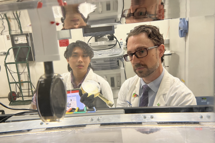
For Fetzer, the most meaningful parts of his career come down to people. “Working with curious, engaged, and hardworking trainees has been extremely rewarding.” He credits students and residents with bringing energy and insight that drive impactful research.
He is equally clear about the role of sonographers. “Ultrasound is incredibly technologist-dependent. I rely heavily on the hard work, dedication, and experience of our sonographers, without whom we would not be able to provide the advanced techniques or high-quality imaging from which our patients benefit.” Many participate directly in protocol development and translational studies, and he considers them colleagues and partners.
His work also depends on partnerships across clinical disciplines. “I have found that the most impactful imaging research occurs with the collaboration, guidance, and support of our colleagues in other clinical departments.” Over time, he has learned that major challenges require collective effort. “No matter how hard a single person works, or how great their ideas are, it’s nearly impossible to tackle truly large or unique problems, much less get them tested and implemented as an impactful tool that requires a large multi-specialty team.”
At UT Southwestern, he found a culture that encourages that kind of growth. “UT Southwestern and the Department certainly put quality and patient care first and foremost.” He describes leaders who support junior faculty development and foster collaboration. “This ‘bottom-up’ approach has certainly facilitated and accelerated my academic progress.”
For Fetzer, academic medicine is defined by forward momentum.
“I don’t see myself as someone who stays still or becomes complacent with how things have always been done, or should be done, but what can be done.”