Search
Research Scientist, Optics, Levi Lab job at UT Southwestern Medical Center, Dallas, TX
https://jobs.utsouthwestern.edu/job/22823923/research-scientist-optics-levi-lab/
Discover Research Scientist, Optics, Levi Lab and other Research & Laboratory jobs in Dallas, TX and apply online today!
A: Cross section showing a vessel (arrow) emerging from the edge of the osseous lamina
https://www.utsouthwestern.edu/edumedia/edufiles/departments_centers/otolaryngology/fig.32.pdf
A: Cross section showing a vessel (arrow) emerging from the edge of the osseous lamina. OC, organ of Corti; ST, scala tympani. B: Scanning micrograph of the under surface of the basilar membrane (BsM) and osseous lamina (OSL). The arrows indicate the spiral vessel beneath the basilar membrane, which is exposed to the perilymphatic space. From Roland PS, Wright C.G. Surgical aspects of cochlear implantation: Mechanisms of insertional trauma. Adv Otorhinolaryngol 2006;64:11-30
Chief Nurse Executive highlights totality of transformative care - Center Times Plus - UT Southwestern
https://www.utsouthwestern.edu/ctplus/stories/2024/state-of-nursing-2024.html
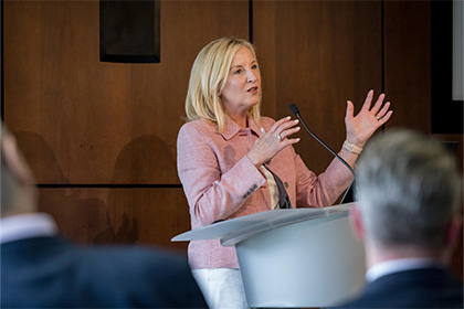
May 9th, UT Southwestern’s annual State of Nursing Address by Associate Vice President and Health System Chief Nurse Executive Susan Hernandez reflected the transformative journey of nursing over the past year.
Cochlear vasculature as Illustrated by Siebenmann (1894)
https://www.utsouthwestern.edu/edumedia/edufiles/departments_centers/otolaryngology/fig.31.pdf
Cochlear vasculature as Illustrated by Siebenmann (1894). The arrow indicates the central auditory vein, which when present, may provide collateral venous drainage for the vessels associated with scala tympani.
Documentary Screening - “The Zebra and The Bear” - UT Southwestern Medical Center
https://engage.utsouthwestern.edu/events/2025/documentary-the-zebra-and-the-bear
The Zebra & The Bear is a documentary film about a mother’s fierce determination to save her daughter from a devastating ultra-rare disease.
Another preparation in which an electrode array was inserted at the time the temporal bone was fresh and unfixed
https://www.utsouthwestern.edu/edumedia/edufiles/departments_centers/otolaryngology/fig.30.pdf
Another preparation in which an electrode array was inserted at the time the temporal bone was fresh and unfixed. After fixation and osmium staining, the array was removed from scala tympani. This SEM image shows the surface of the spiral ligament in an area where the array contacted the lateral wall. In the upper portion of the image the spiral ligament tissue is seen to be compressed and marked by grooves produced by the electrode. Below that area the normal porous surface of the spiral
This is a temporal bone which had an implant electrode inserted at the time that the bone was fresh and unfixed
https://www.utsouthwestern.edu/edumedia/edufiles/departments_centers/otolaryngology/fig.29.pdf
This is a temporal bone which had an implant electrode inserted at the time that the bone was fresh and unfixed. After fixation, osmium staining, and dissection the electrode was removed from scala tympani. “A” shows the apical turn in an area that the electrode did not reach. The arrows indicate normal-appearing venules in the lower part of the spiral ligament. “B” shows an area in the basal turn where the electrode was in contact with the lateral wall immediately below the basilar
Veterinary Medical or Radiation Oncologist job at UT Southwestern Medical Center, Dallas, TX
https://jobs.utsouthwestern.edu/job/22868299/veterinary-medical-or-radiation-oncologist-dallas-tx/
Discover Veterinary Medical or Radiation Oncologist and other Faculty and Physicians jobs in Dallas, TX and apply online today!
Cochlear dissection after insertion of an implant electrode array
https://www.utsouthwestern.edu/edumedia/edufiles/departments_centers/otolaryngology/fig.28.pdf
Cochlear dissection after insertion of an implant electrode array. Portions of the osseous lamina and basilar membrane have been removed to more clearly show the electrode lying in scala tympani. The silicone carrier of the array has been lightly stained with osmium so that it has a light brown color. Note that in the area indicated by the arrows the array is in direct contact with the lateral wall under the basilar membrane. From Roland PS, Wright C.G. Surgical aspects of cochlear
UT Southwestern No. 1 hospital in Dallas-Fort Worth, Best Hospital rankings show: Newsroom - UT Southwestern, Dallas, Texas
https://www.utsouthwestern.edu/newsroom/articles/year-2022/july-us-news-best-hospital.html?utm_source=facebook&utm_medium=organic&utm_campaign=newsroom&utm_content=usnwrrankings&utm_utm-audience=na&fbclid=IwAR3tY4r24j2gT-WKC5ar4jpT51bBLwVrirgZfpXOMV_c4aSikMhT6vdPJaI
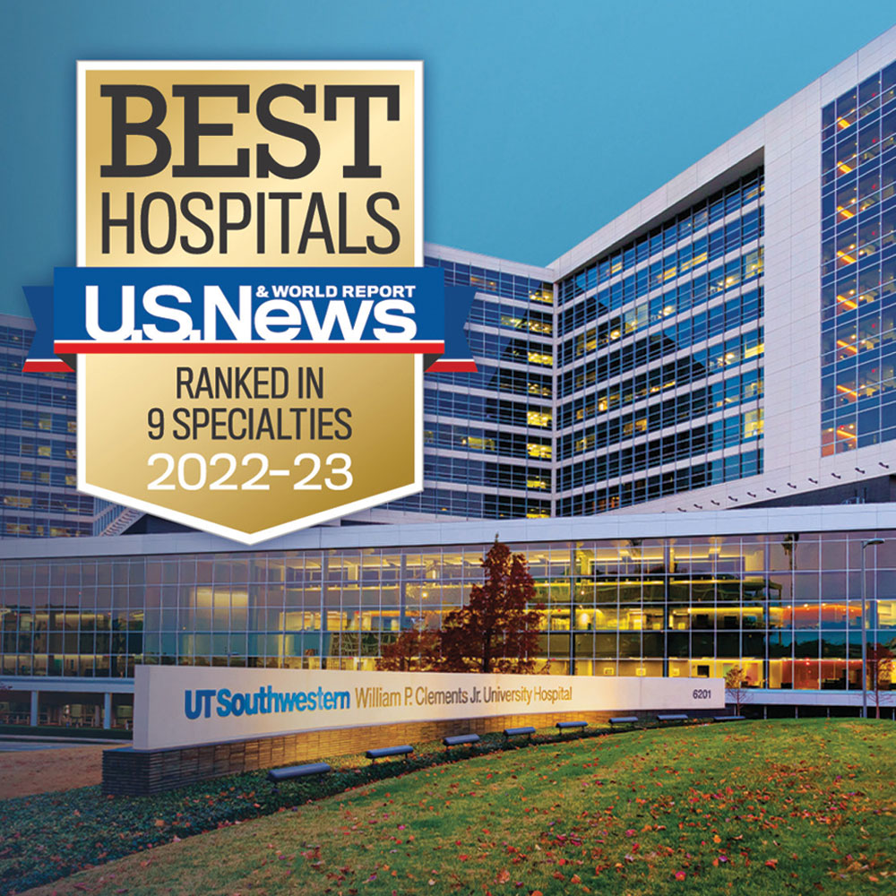
UT Southwestern Medical Center is the No. 1 hospital in Dallas-Fort Worth – the nation’s fourth-largest metro area – for the sixth consecutive year and ranks among the top hospitals nationally in nine specialties ranging from brain to heart to cancer care.