Search
Office of Academic and Physician Recruitment: UT Southwestern, Dallas, TX
https://www.utsouthwestern.edu/about-us/administrative-offices/academic-recruitment//
Office of Academic Recruitment Support Services partners with departments, centers, and leadership to support the recruitment, onboarding, and retention efforts of faculty.
Training Verification Requests: Emergency Medicine - UT Southwestern, Dallas, Texas
https://www.utsouthwestern.edu/departments/emergency/education/training-verification/
Training Verification Requests Verification of Graduate Medical Education Training To request verification of residency or fellowship training, please complete the verification request form . Back-to top
Submissions - Forefront - UT Southwestern Medical Center
https://engage.utsouthwestern.edu/forefront/submissions
Share your updates and achievements for publication in an upcoming issue of Forefront, UT Southwestern's monthly digital publication for alumni.
Heitman, Wang elected as AAAS Fellows - CT Plus - UT Southwestern
https://www.utsouthwestern.edu/ctplus/honors-awards/heitman-wang-aaas-2024.html
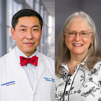
Elizabeth Heitman, Ph.D., Professor in the Department of Psychiatry, and Andrew Wang, M.D., Professor of Radiation Oncology, have been elected as Fellows of the American Association for the Advancement of Science (AAAS).
Corporate Mentoring: WISMAC - UT Southwestern, Dallas, TX
https://www.utsouthwestern.edu/edumedia/edufiles/life_at/wismac/corporate-mentoring.pdf
* * * Some would believe that it’s simply not possible to be successful because of various obstacles that will get in their way, like the organizational culture or their own work-life priorities * The fact that in most organizations there are few female role models at the top serves as prima facie evidence that the opportunities for women are limited. The natural assumption when looking up the organization is that others have tried and not succeeded and “my chances aren’t good
633260Spring-2011-ADC-Newsletter.pdf
https://www.utsouthwestern.edu/media/633260Spring-2011-ADC-Newsletter.pdf
On Jan. 4, 2011, President Obama signed into law the National Alzheimer’s Project Act (NAPA). Thousands of Americans from every walk of life sent messages to Congress and the White House to urge its passage. All of us in the Alzheimer’s Disease Center at UT Southwestern strongly support- ed this initiative and worked closely with the Alzheimer’s Association to lobby legislators. NAPA’ s objective, the associa- tion says, is “to create a coordi- nated national plan to overcome
Southwestern Sweethearts - Bethany and Mack Grubb - UT Southwestern Medical Center
https://engage.utsouthwestern.edu/pages/stories/southwestern-sweethearts---bethany-and-mack-grubb
story Valentine's Day
Looking back and moving forward with the Chief Nurse Executive: Center Times Plus, UT Southwestern
https://www.utsouthwestern.edu/ctplus/stories/2025/nursing-2025.html
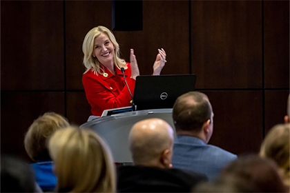
State of Nursing address during National Nurses Week gathered the UTSW nursing community to reflect on the past year, recognize progress, and look ahead to the future of nursing across the Health System.
Southwestern Sweethearts - Drs. English - UT Southwestern Medical Center
https://engage.utsouthwestern.edu/pages/stories/southwestern-sweethearts---drs.-english
story Valentine's Day
MSTLC.pdf
https://www.utsouthwestern.edu/edumedia/edufiles/departments_centers/neurology/MSTLC.pdf
Team TLC Patients at UT Southwestern’s multiple sclerosis center receive more than just cutting-edge medical care. They receive help in living better lives while they battle their debilitating disease. T o t a l L i f e C a r e Susan Sides adored helping patients as a nurse at Children’s Medical Center Dallas. She held increas- ingly responsible positions there for more than 15 years after her first episode of multiple sclerosis and even after a 2002 stroke, all that time