Search
Dissection in which the osseous lamina and basilar membrane have been removed so as to show the modiolar wall in the basal cochlear turn of an osmium-stained specimen
https://www.utsouthwestern.edu/edumedia/edufiles/departments_centers/otolaryngology/fig.13.pdf
Dissection in which the osseous lamina and basilar membrane have been removed so as to show the modiolar wall in the basal cochlear turn of an osmium-stained specimen. Neural elements associated with the modiolar wall are well displayed. “G” indicates the spiral ganglion. Peripheral (P) and central (C) processes of spiral ganglion cells are seen emerging from the ganglion along the length of the basal turn. ST, floor of scala tympani. This preparation shows the same anatomical features
Cochlear dissection after insertion of an Advanced Bionics Thin Lateral electrode array designed to track the lateral wall of scala tympani
https://www.utsouthwestern.edu/edumedia/edufiles/departments_centers/otolaryngology/fig.9.pdf
Cochlear dissection after insertion of an Advanced Bionics Thin Lateral electrode array designed to track the lateral wall of scala tympani. With this array, contacts are placed immediately beneath the basilar membrane. From Wright, CG, Roland PS, Kuzma J. Advanced Bionics Thin Lateral and Helix II electrodes: a temporal bone study. Laryngoscope 115(11);2005:2041-2045. Copyright Lippincott, Williams and Wilkins. Reproduced with permission. http://lww.com
Cochlear dissection after insertion of a cochlear implant electrode array (Advanced Bionics Thin Helix)
https://www.utsouthwestern.edu/edumedia/edufiles/departments_centers/otolaryngology/fig.8.pdf
Cochlear dissection after insertion of a cochlear implant electrode array (Advanced Bionics Thin Helix). The array has been colored blue to provide better contrast of the silicon carrier. The array lies in scala tympani and is situated beneath the translucent basilar membrane. The apical cochlear turn has been removed to provide an unobstructed view of the basal turn. From Wright, CG, Roland PS, Kuzma J. Advanced Bionics Thin Lateral and Helix II electrodes: a temporal bone study
Osseous lamina with its nerve fibers in a specimen oriented in nearly horizontal plane
https://www.utsouthwestern.edu/edumedia/edufiles/departments_centers/otolaryngology/fig.7.pdf
Osseous lamina with its nerve fibers in a specimen oriented in nearly horizontal plane.
A preparation in which the modiolus and osseous lamina are seen in profile after removal of the otic capsule bone covering the human cochlea
https://www.utsouthwestern.edu/edumedia/edufiles/departments_centers/otolaryngology/fig.6.pdf
A preparation in which the modiolus and osseous lamina are seen in profile after removal of the otic capsule bone covering the human cochlea.
Cross section of mouse cochlear duct, basal turn, showing internal structure of organ of Corti to complement surface views seen in the previous images
https://www.utsouthwestern.edu/edumedia/edufiles/departments_centers/otolaryngology/fig.5.pdf
Cross section of mouse cochlear duct, basal turn, showing internal structure of organ of Corti to complement surface views seen in the previous images.
Higher power view of osseous lamina and basilar membrane/organ of Corti in a preparation from an elderly individual in which there has been mild thinning of the myelinated nerve fibers within the osseous lamina
https://www.utsouthwestern.edu/edumedia/edufiles/departments_centers/otolaryngology/fig.3.pdf
Higher power view of osseous lamina and basilar membrane/organ of Corti in a preparation from an elderly individual in which there has been mild thinning of the myelinated nerve fibers within the osseous lamina. The spirally oriented nerve fibers in the osseous lamina are nicely displayed in this specimen.
A: Otic capsule bone drilled to a thin shell to reveal contours of the cochlea
https://www.utsouthwestern.edu/edumedia/edufiles/departments_centers/otolaryngology/fig.1.pdf
A: Otic capsule bone drilled to a thin shell to reveal contours of the cochlea. The membranous labyrinth has been stained with osmium so that it appears dark through the thinned bone. An implant electrode array had been inserted into this specimen and a portion of the array can be seen at lower left. B: Similar preparation in which the bone overlying scala vestibuli has been removed to show the osmium-stained basilar membrane and osseous spiral lamina.
COVID-19 vaccination info for 2024-25
https://www.utsouthwestern.edu/employees/campus-updates/articles/year-2024/covid-shots-2024.html
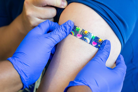
COVID-19 vaccination info for 2024-25
Human Resources Info for FY26
https://www.utsouthwestern.edu/employees/campus-updates/articles/year-2025/hr-changes-fy26.html

hr human resources updates fiscal year 2026