
Optical and Ultrasound Images
Here are some optical and ultrasound images from the Small Animal Imaging Resource at UT Southwestern (UT-SAIR).
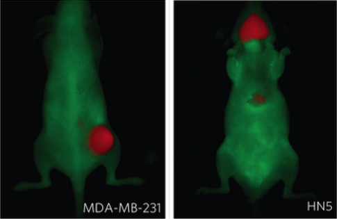
Fluorescent Imaging Using CRi Maestro. Ultra pH-sensitive fluorescent nanoprobes for cancer detection. Novel pH sensitive nanoprobe for detecting tumors growing in mice (breast and brain cancers shown). Further details in “A nanoparticle-based strategy for the imaging of a broad range of tumours by nonlinear amplification of microenvironment signals.“ Wang, Zhou, Huang, Hensley, Huang, Ma, Zhao, Sumer, DeBerardinis, Gao, Nature Materials, 13 (2014) 204-212.
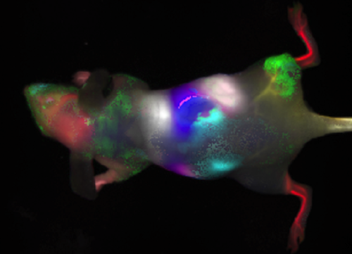
Dynamic Fluorescent Imaging of ICG Infusion Using CRi Maestro. Infusion of indocyanine green (ICG) in the tail vein of nude mouse with MCF7 human breast tumor xenograft in right thigh. Principal component analysis of the signal intensity variation following ICG infusion (50 µl, 260 µM) allows identification of organs based on perfusion characteristics (DyCE analysis). Further details in J. Biomed. Nanotechnol. 10, 1–8, 2014.
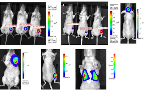
Detection of Diverse Breast Tumors Using IVIS Spectrum or Lumina. Each image shows bioluminescent image acquired within 15 minutes following administration of luciferin subcutaneously in foreback neck region of mouse. Top left: MDA-MB231-luc in SCID mice (no depilation required). Top center MDA-MB231-luc in nude mice. Top right: MDA-MB231-HRE-luc implanted in brain and observed through intact skull. Bottom left: orthotopic syngeneic 4T1-luc in mammary fat pad; bottom center: MCF7-luc growing subcutaneously in nude mouse thigh. Bottom right MCF7-luc growing in lungs following IV injection in tail vein of nude mouse.
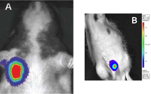
BLI Detection of Tumors in Rats Using IVIS Spectrum or Lumina. Left: orthotopic A549-luc tumor growing in lungs of nude rat following direct injection of cells. Further details in Saha, Watkins, Yin, Thorpe, Story, Song, Raghavan, Timmerman, Chen, Minna and Solberg, RADIATION RESEARCH 174, 62–71 (2010). Right: Examination of orthotopic PC3- luc prostate tumor in adult male nude rat at day 9 after tumor implantation using in vivo bioluminescence imaging: rat was anesthetized with 2.0% isoflurane in oxygen D-luciferin (500 μl of 150 mg/kg sodium salt in saline) was injected SQ in foreback neck and BLI signal observed 10 minutes after injection using IVIS® Lumina Imaging System. Data acquired in association with Biomedical Engineering Graduate Student Derek White, supported by R01 CA139043.
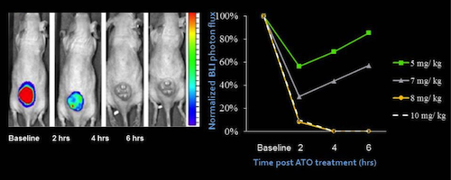
Optical Assessment of Vascular Disruption in U87-mCherry-luc Tumors Using IVIS Spectrum or Lumina: Optical imaging was performed at various times before and after administration of ATO. On each occasion FLI was performed first and then D-luciferin was administered SC in the neck and BLI was performed over a period of 16 minutes. Left: Sequential BLI at the 10 minute time point following luciferin administration with respect to a dose of 8 mg/kg ATO administered IP. Right: Normalized BLI signal intensity at 10 minute time point after administration of luciferin indicating vascular shutdown following various doses of ATO. Further details available at doi:10.1371/journal.pone.0046106.g002
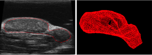
High Resolution Ultrasound of SC Tumor Rat Prostate Tumor: B mode image and wire mesh rendering from 3D data set obtained in B-mode using Vevo770.
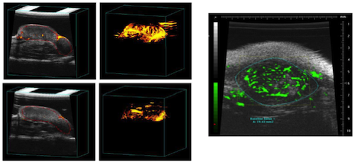
Vascular Assessment Using Ultrasound (Images acquired using Visual Sonics Vevo770). Left: Vascular disruption assessed by power-Doppler Ultrasound (PD-US) in MCF7-mCherry-luc tumors with respect to vascular disruption caused by arsenic trioxide (ATO; 8 mg/kg). PD US images are presented as single slice (left) and PD maximum intensity projection (right) before and 4 hours after treatment with ATO. Further details in doi:10.1371/journal.pone.0046106.g004. Right: High resolution ultrasound using a VisualSonics Vevo770 small animal ultrasound device of an 8 mm diameter syngeneic MTLn3 mammary tumor growing subcutaneously in a Fisher rat. In this case Doppler mode using 40 MHz transducer showed only a few major blood vessels in tumor, but following infusion of commercial contrast microbubbles extensive vasculature was observed. Further details in Integr. Biol., 3, 375-387, 2011 DOI: 10.1039/C0IB00135J; PMID: 21321746.
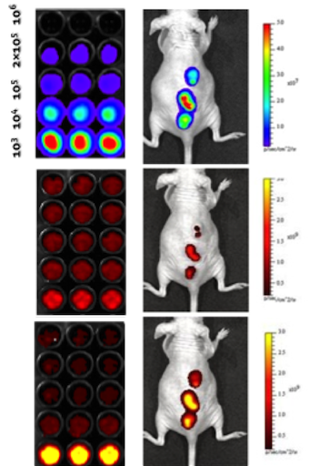
Multimodal optical imaging of 9L-luc-GFP-mCherry cells in vitro and tumors in vivo using IVIS Spectrum or Lumina. Left: Imaging of various numbers of 9L-luc-GFP-mCherry cells in well plate by fluorescence (mCherry λ ex 570nm λ em 610nm (bottom) and GFP λ ex 430nm λ em 465nm (middle)) and BLI following addition of luciferin (top). Right: Images of mCherry fluorescence (bottom), GFP fluorescence (middle) and BLI following administration of luciferin (top) of mouse with three triply transfected tumors growing on back of nude mouse. Data acquired by Dr. Li Liu.
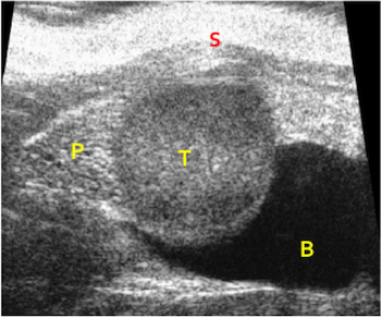
High resolution ultrasound of orthotopic prostate tumor. Obtained in B-mode using Vevo770: T tumor; P normal prostate; B bladder; S skin.