

UTSW researchers gain powerful new tools to explore gene expression cell by cell and in the spatial context of the tissue
During transcription, the information encoded within the DNA sequence of a gene is copied into a strand of RNA, called an RNA transcript. The transcriptome – the population of RNA transcripts within a cell – provides a picture of active gene expression and biological pathways within a cell.
Bulk RNA sequencing is a commonly used method to study the gene expression signature of a given tissue. Since tissue is comprised of more than one cell type, this sequencing method provides an “average” gene expression profile based on the population of cells present in that tissue specimen. But this technique is limited in assignment of a gene signature to a particular cell type.
“Now, with the advent of the single-cell RNA sequencing method, researchers can study gene expression at the individual-cell level, enabling precise mapping of cell type and state specific gene signatures in the context of health and disease,” said Prithvi Raj, Ph.D., Associate Professor of Immunology and Director of the Microbiome Research Laboratory, Genomics Core facility, and Microarray and Immune Phenotyping Core facility at UT Southwestern.
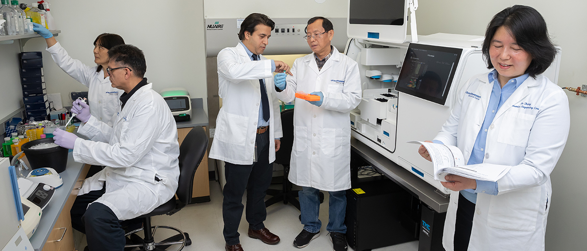
Since it was established in 2001, the Immunology (Genomics and Microarray) Core has been providing diverse genomics and proteomics services to hundreds of UTSW investigators working on basic science and clinical research projects. In 2018, the Microbiome Core was added to provide microbial genomics services to characterize microbiome, bacterial, fungal, and viral pathogens using next-generation sequencing technology.
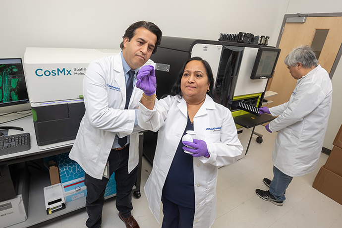
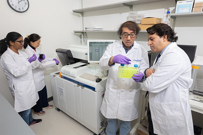
To help researchers stay at the forefront of scientific advances, the Immunology Core recently acquired advanced instrumentation from NanoString Technologies and 10x Genomics. These cutting-edge platforms enable quantitative analysis of gene expression in individual cells and in the spatial context of tissues. According to Dr. Raj, they offer more comprehensive, multimodal information about changes in gene expression compared with bulk RNA sequencing methods.
Following genes and their changes
Spatial transcriptomics is a technique that allows researchers to measure gene expression at specific locations within a tissue specimen. It creates a map of genes expressed within the tissue’s native structure, providing insights into cellular interactions and tissue organization at a molecular level.
Using spatial transcriptomics, researchers can illuminate differential gene signatures between a tumor and normal site in the same tissue section, providing insight on molecular changes involved in normal cells turning cancerous, for example. This technology also can capture cross talk between healthy and cancerous cells as well as within the local microenvironment.
“Now, with the new instruments, we can precisely study gene expression signatures at single cell or subcellular resolution and gain insight into disease-driving mechanisms,” Dr. Raj said. “For example, you can ask which cell types are involved in pathogenesis, progression, and treatment responses.”
The Immunology Core houses these two new spatial transcriptomics platforms: Visium HD/Xenium from 10x Genomics and GeoMx/CosMx from NanoString Technologies. These machines can capture information on the entire transcriptome or a targeted set of up to 6,000 genes. Currently, only a few of these machines are in use in Texas.
One platform, Chromium X from 10x Genomics, uses microfluidic technology to isolate individual cells into tiny oil droplets, enabling high throughput single-cell analysis. Within each droplet, the platform sequences the transcriptome of a single cell, allowing researchers to profile gene expression, immune repertoire, and other epigenetic changes in thousands of single cells.
The Immunology Core at the BioCenter provides consultation for study design, sample collection, and data generation services to investigators, Dr. Raj said.
Technology used to study tendon recovery
Robert Tower, Ph.D., an Assistant Professor of Surgery at UTSW who focuses on musculoskeletal development and regeneration, began using single-cell and spatial transcriptomics as a postdoctoral researcher at the University of Pennsylvania and Johns Hopkins University.
Dr. Tower and his colleagues used single-cell and spatial transcriptomics to study regeneration of injured tendons. Using research tools at the Microbiome Core facility, they found that sensory-nerve growth was essential for proper tendon recovery. His team also used the techniques to study skull development, which begins with small islands of bone that grow and fuse together. “How fast they fuse is really important, and working with the Core facility, we found that this is regulated by sensory nerves,” he said.
“The use of this technology is what attracted me to UT Southwestern,” Dr. Tower added. “We’ve studied long bones, fractures, and neurofibromatosis. People could think this technology is just the ‘new hot thing,’ but a lot of the time this work is not possible without spatial information.”
For more information on the Immunology Core facility (Genomics, Microarray and Microbiome) and its services, call 214-648-7324 or contact Dr. Raj via Prithvi.raj@utsouthwestern.edu.