

New insights into how cells target mitochondrial destruction could hold treatment potential
Mitochondria – the “powerhouses of the cell” – require constant vigilance. When they’re malfunctioning or no longer needed, the cells they inhabit target and destroy them. But UT Southwestern researchers have discovered that mitochondria have a way to limit this destruction to the specific area that needs repair.
And because mitochondria affect so many bodily functions and diseases – including neurodegeneration, cancer, and fatty liver disease – understanding mitochondrial repair and how to boost or suppress it could pay off someday in treating diseases.
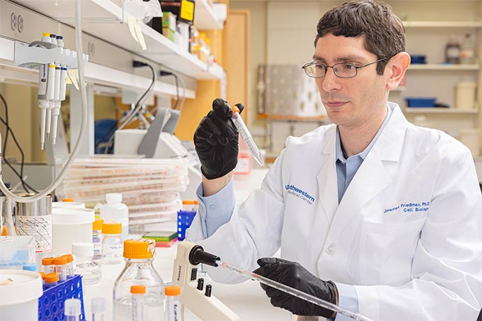
“It’s just like a car – when you get wear and tear, you want to repair the damaged part rather than replace the whole thing,” said Jonathan Friedman, Ph.D., Assistant Professor of Cell Biology and senior author of the study, which was published online by the Journal of Cell Biology.
Dr. Friedman usually studies mitochondrial processes and their relation to cristae, the interior folds that house the energy factories. His lab focuses on how cristae membranes can shape and reshape themselves according to the needs of the cell.
His team wasn’t expecting to get caught up in mitophagy (“mitochondrion-eating”), a set of processes that cells use to destroy mitochondria, when they began their experiments. They were instead investigating a protein complex called MICOS that helps cristae maintain their shape.
“Some of the most fun we have as scientists is when we start looking for one thing and find another,” Dr. Friedman said. “Our lab didn’t really study mitophagy going in – we had to learn the best ways to look at it.”
Using cultured human cells to track the biochemical pathways that MICOS is involved in, they discovered that a protein called TMEM11 links the MICOS complex to another set of proteins involved in mitophagy.
The process was tricky to follow because when cells are healthy, mitophagy proteins all exist in very low numbers, Dr. Friedman explained.
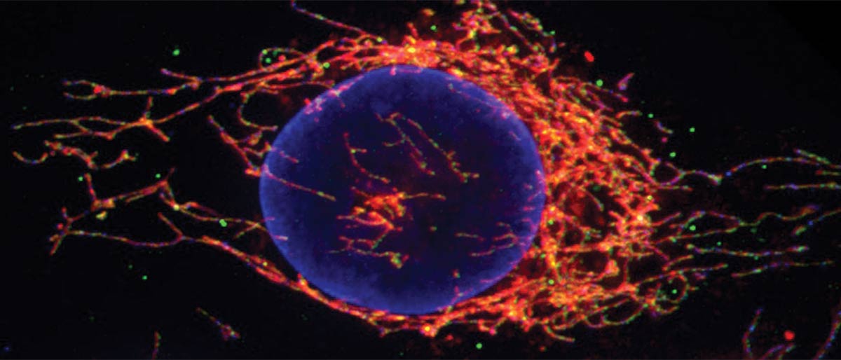
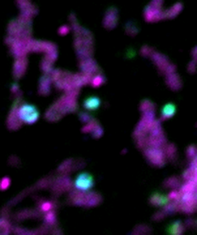
But graduate student and study first author Mehmet Oguz Gok, using a technique called confocal fluorescence microscopy, saw TMEM11 and the mitophagy protein BNIP3 go to the same place at the same time when the mitophagy pathway was activated.
“Oguz was extremely excited when he saw the images – I think he knew even faster than I did how critical that result was,” Dr. Friedman said.
When the researchers genetically manipulated cells to block TMEM11, mitophagy became hyperactivated and occurred at many more places at once, meaning that TMEM11 might be helping to limit the site of the removal to the defective parts of mitochondria.
“Now that we know this process can be spatially controlled, we want to know how the cell knows there’s something wrong with mitochondria at a particular spot and needs to be destroyed,” Dr. Friedman said.
Dr. Friedman’s team collaborated with the lab of Prashant Mishra, M.D., Ph.D., Assistant Professor at the Children's Medical Center Research Institute at UT Southwestern (CRI), to monitor mitochondrial function in cells without TMEM11. Both Drs. Friedman and Mishra are members of the Harold C. Simmons Comprehensive Cancer Center.
An exciting aspect of the work is that TMEM11’s activity provides a clue on how to manipulate mitophagy as a tool to promote overall mitochondrial health. Mitochondrial dysfunction is commonly associated with aging and underlies many diseases. Therefore, finding ways to promote mitophagy may have therapeutic potential.
Other researchers involved in the study were Olivia Connor, Research Technician; Xun Wang, Ph.D., Assistant Instructor at CRI; Cameron Menezes, a student in the Medical Scientist Training Program; and Claire Llamas, Senior Research Associate at CRI. The work was supported by the National Institutes of Health (1S10OD021685-01A1, P30CA14253, 1S10OD028630-01, R00HL133372, R35GM137894, 1DP1ES030449, and 1R01AR073217) and the UT Southwestern Endowed Scholars Program.
Dr. Friedman is a Southwestern Medical Foundation Scholar in Biomedical Research.