

Advanced microscopy reveals how cholesterol crystallizes into a more dangerous form
UTSW researchers may have solved the long-standing mystery of the genesis of crystallized cholesterol, a form considered more likely to clog the arteries in humans. Their study, published recently in the Journal of Cell Biology, points to a novel therapeutic target that could eventually lead to new ways to treat or delay the development of cardiovascular diseases.
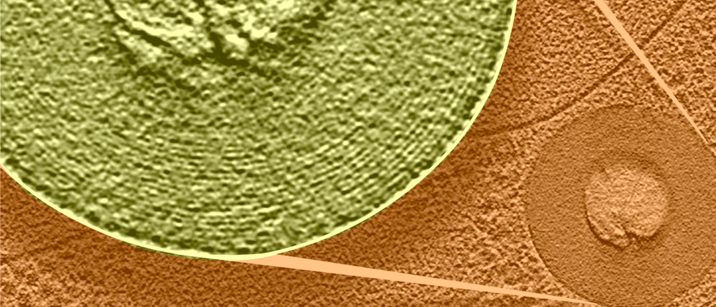
Using advanced microscopy, the researchers observed lipid droplets in yeast cells undergoing phase transitions, transforming from disordered molecules to a lipid-crystalline lattice state in response to glucose starvation.
Atherosclerosis, a buildup of fatty plaques in the artery walls, increases the risk of heart attack, stroke, and congestive heart failure. “Crystallized cholesterol deposits had been observed for decades, but how they formed in cells was poorly understood,” said Mike Henne, Ph.D., Associate Professor of Cell Biology and Biophysics. “Because yeast is a model organism for mammalian cells, the same crystallization process likely occurs in humans.”
He added that working to understand this change of state is an emerging field of study. “Although scientists have observed cholesterol crystallizing in cells when large amounts of cholesterol are added to a lab dish, our study is the first to our knowledge to thoroughly determine the trigger and mechanism for that change, and to do so in cells. Glucose starvation is the trigger, and the breakdown of triglyceride to free fatty acids that the cell can use as a backup energy source appears to be the mechanism driving crystallization,” Dr. Henne said.
Strikingly, the crystallization process appears to change the population of proteins that rest on the surface of lipid droplets, very likely affecting the way these organelles interact and function within the cell – a finding that generates new questions for future studies, the researchers said.

Dr. Henne is a corresponding author of the study along with Daniela Nicastro, Ph.D., Professor of Cell Biology and an expert in the cryo-electron tomography (cryo-ET) technology that made it possible to view the cells in a life-like state.
Cryo-ET is a cryo-electron microscopy (cryo-EM) method that provides three-dimensional images of life-like preserved cells, featuring subcellular structures at molecular resolution. Powerful computers then reconstruct 3D-cellular structures. To preserve their native structures during imaging, the biological samples are flash-frozen and imaged at cryogenic temperatures (at about -196 degrees Celsius).
Dr. Nicastro, who was instrumental in setting up UT Southwestern’s cryo-EM center, is advancing a technique called cryo-FIB (cryo-focused ion beam milling) that was key in this study. “One challenge we faced was that budding yeast cells are 50 times thicker than what can be imaged directly by cryo-EM,” she said. “Therefore, after rapidly freezing the yeast cells, we had to ‘section’ them. We used a very finely focused beam of gallium ions to mill away material from the top and bottom of each cell, leaving behind a thin lamella, or section, that was suitable for imaging.”
Dr. Henne said next steps in the investigation include screening for drug candidates and investigating whether the same phenomenon happens in human cells.

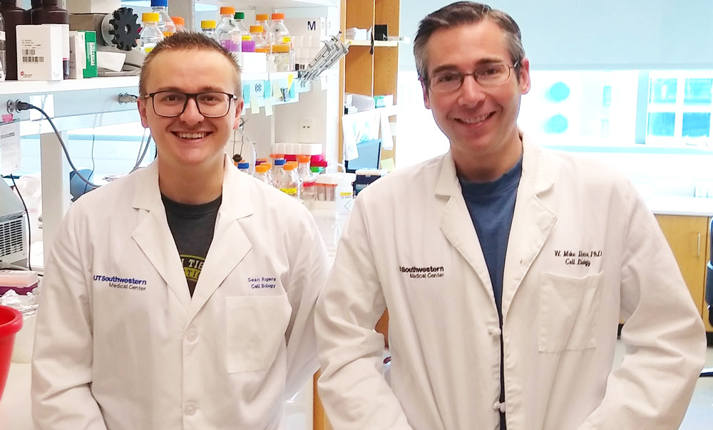
Lead authors were Sean Rogers, Ph.D., a former Henne lab graduate student now at Washington University in St. Louis, and Long Gui, Ph.D., a Research Scientist in the Nicastro lab. Researchers at the Sorbonne/University of Paris, and the University of Fribourg in Switzerland also participated.
The study received support from the National Institutes of Health, The Welch Foundation, the Ara Parseghian Medical Research Fund, the UT Southwestern Endowed Scholars Program in Medical Science, the Cancer Prevention and Research Institute of Texas, supporting UTSW’s Cryo-EM Microscopy Facility. The Swiss National Science Foundation, the Swiss National Supercomputing Center, and the French National Research Agency also provided support.
More information on this research, including the full list of authors, can be found in the study.
Dr. Henne is a W.W. Caruth, Jr. Scholar in Biomedical Research.