

President’s Lecture Series: The promise of cryo-electron microscopy
Microscopes that can look inside human cells and create 3D images of objects as tiny as a ribosome – with details all the way down to atomic level – are now a reality.
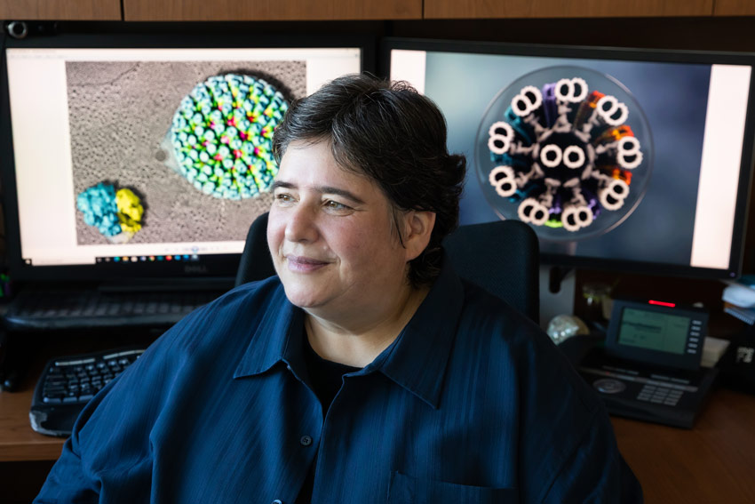
Daniela Nicastro, Ph.D., Professor of Cell Biology, helped plan and direct UT Southwestern’s Cryo-Electron Microscopy Facility, which opened in 2016 with microscopes that can accomplish such feats. At 4 p.m. on Thursday, Oct. 28, she will give a virtual talk titled “Cryo-Electron Microscopy: Opening a Frosty Window into Cells” as the next speaker in the President’s Lecture Series. Register here for the event.
Dr. Nicastro will describe how cryo-electron microscopy (cryo-EM) works and discuss the ways this technology has allowed scientists to uncover more information about the function of proteins and small organelles to gain a greater understanding of various cellular processes. Because cryo-EM specimens are flash-frozen with the water inside retained – rather than dried out like a raisin and preserved in resin – scientists now get a more life-like image, she explained.
“Using cryo-electron tomography, a specialized cryo-EM technique, we can visualize the 3D structure of proteins and macromolecular complexes inside cells,” Dr. Nicastro said. “By observing these components in their natural cellular environment, we learn about their distribution within the cell and how they interact with each other and function.
“This will ultimately provide a better understanding of the inner workings of healthy cells and can reveal what goes wrong on a cellular level in diseases,” she added. “Seeing is understanding.”
In cryo-electron microscopy, specimens are rapidly frozen to prevent the formation of damaging ice crystals and to retain their natural water. They are kept frozen at cryogenic temperatures as they are viewed, which helps protect them from damage by the microscope’s high-energy electron beam and high vacuum – while allowing them to be seen in their near-native state.
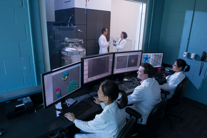
The electron microscope then creates a high-resolution image by bombarding specimens with fast-moving electrons, resulting in dark or light areas, depending on whether the electrons pass through a relatively empty space or get blocked by material in the specimen. The results are then processed in computers to generate 3D images.
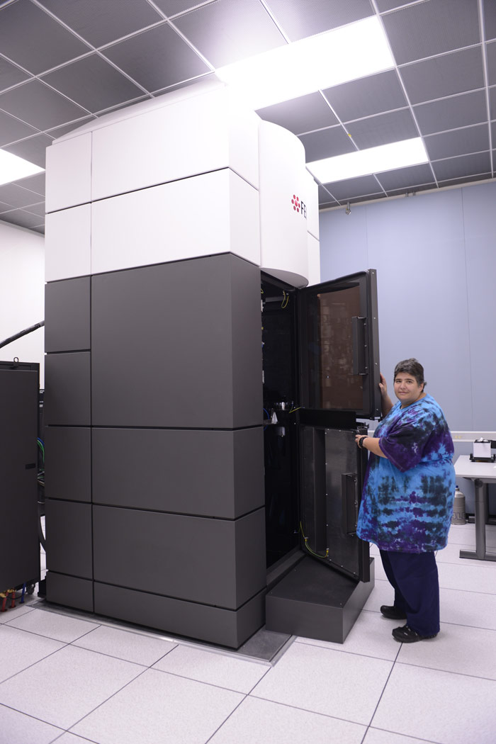
At UT Southwestern’s cryo-EM facility, with locations on North and South Campus, powerful cryo-electron microscopes called Titan Krioses stand about 12 feet tall and weigh 2 tons apiece. The microscopes, one at each site, can keep specimens at minus 321 degrees. Two additional, slightly smaller, cryo-electron microscopes are also available for researchers, as well as an Aquilos 2 machine used to precisely carve thin slices of cells to be imaged. These instruments place UT Southwestern among the top institutions in the world for cryo-EM, said Dr. Nicastro.
Her journey to becoming a cryo-EM expert at UT Southwestern began as undergraduate – a zoologist in training – studying the evolution of sensory neurons in the antenna of mosquito and fly larva in her native Germany. Now she peers deeper inside cells to study the structure and function of molecules using UT Southwestern’s instruments.
Dr. Nicastro works to continue development of cryo-EM and imaging techniques here. She also does research on the internal skeleton (or cytoskeleton) of cells, and the hair-like appendages on the cell surface called cilia and flagella that help cells move and sense their environment. Another area of interest of her lab is host-pathogen interactions, such as how parasites and viruses invade host cells.
One of her most intriguing findings using the cryo-EM technology, she said, was watching how the thousands of tiny motors on a single cilium are coordinated in a “synchronized dance” that leads to the undulating motion of a cilium that propels a cell.
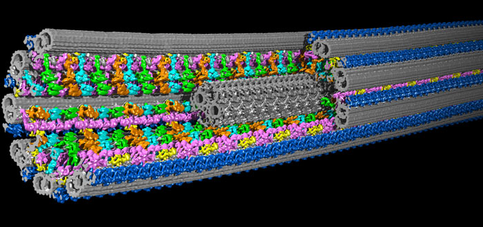
Credit: Dr. Daniela Nicastro
A study using cells from a patient led to a report this year in Molecular Biology of the Cell that describes structural defects in cilia present in people with primary ciliary dyskinesia (PCD), a genetic disease that makes it difficult for sufferers to clear mucus properly from their respiratory system, leading to chronic lung, ear, and sinus infections, along with other disorders.
Dr. Nicastro, a Cancer Prevention and Research Institute of Texas (CPRIT) Scholar, came to UT Southwestern in 2015 from Brandeis University, where she was Associate Professor of Biology.
She received her Diplom and Ph.D. in biology from the Ludwig-Maximilians University in Munich, Germany, and did postdoctoral research in structural biology at the Max Planck Institute of Biochemistry, also in Munich, and in structural cell biology at the University of Colorado Boulder.
Dr. Nicastro has been named a Pew Biomedical Scholar, a Keith R. Porter Fellow, and received the 2020 WICB (Women in Cell Biology) Mid-Career Award for Excellence in Research from the American Society for Cell Biology.