

Simmons Cancer Center investigators receive more than $5 million in CPRIT funding
The Harold C. Simmons Comprehensive Cancer Center at UT Southwestern received more than $5 million in research funding in the latest round of grants awarded by the Cancer Prevention and Research Institute of Texas (CPRIT).
“Texas voters strongly reaffirmed their support for CPRIT in last year’s election, with nearly two-thirds of them approving another $3 billion in bonds to fund cancer research and prevention. The research work continues apace with new funding that will advance the Simmons Cancer Center’s work in immunotherapy, radiation oncology, and the development of new blood tests,” said Dr. Carlos L. Arteaga, Director of the Simmons Cancer Center.
Liver cancer screening
Screening for liver cancer is the focus of Dr. Amit Singal, Professor of Internal Medicine and Population and Data Sciences, Medical Director of the Liver Tumor Program, and Clinical Chief of Hepatology at UT Southwestern. He received a $2.5 million CPRIT grant that builds on previous funding from CPRIT and the National Cancer Institute.

His work seeks to improve screenings for liver cancer. Current screening methods, including ultrasounds and a serum biomarker, miss more than a third of early detections even though the ultrasounds are conducted as frequently as every six months for patients at higher risk because of cirrhosis. Patients with late-stage detection of liver cancer have a median survival of less than a year, so early diagnosis is critical.
The CPRIT funding will allow Dr. Singal to further test a new blood biomarker that shows promise in detecting liver cancer and is expected to be superior to a serum biomarker currently in use. Dr. Singal will also use artificial intelligence to analyze medical records of patients with cirrhosis and stratify them based on risk.
“Overall, these data would have the potential to immediately and radically transform our approach to liver cancer risk assessment and early detection in patients with cirrhosis, thereby taking the first big step to reducing liver cancer mortality,” he said.
Dr. Singal discloses that he serves as a consultant to Glycotest, which developed the blood panel he is studying.
Immunotherapy for liver cancer
Dr. David Hsieh, Assistant Professor of Internal Medicine, is studying immunotherapy for liver cancer. He received nearly $1.5 million to investigate whether a drug may enhance the effectiveness of existing immunotherapies for liver cancer. The drug, a type of sulfonamide called E7820, was previously tested in patients as an anti-cancer drug but was not found to be broadly effective.
Dr. Hsieh’s proposal that E7820 might be able to kick-start immunotherapy and his work on the next steps is an example of collaboration across labs at UT Southwestern. The precise mechanism of E7820 was discovered by Dr. Deepak Nijhawan, Associate Professor of Internal Medicine and Biochemistry, and Dr. Hsieh joined his lab as a fellow in 2018. In the Nijhawan lab, Dr. Hsieh worked with colleagues to develop biomarker tests to determine whether E7820 may kill blood cancers. However, because E7820 induces alternative splicing, this drug may also be used to make cancer cells more recognizable to the immune system. Unmasking cancer so the immune system can see it is a crucial step in getting immunotherapy to unleash its cancer-killing potential.
Dr. Hsieh collaborated with Dr. Hao Zhu, Associate Professor of Internal Medicine at the Children’s Medical Center Research Institute at UT Southwestern and of Pediatrics, to test the drug combined with immunotherapy in animals. He said his CPRIT grant will complete the animal trials and start clinical trials in patients with liver cancer. Dr. Hsieh does not have any relationship with companies that developed E7820.
Immunotherapy for lung cancer
Dr. Kathryn O’Donnell, Associate Professor of Molecular Biology, received a $900,000 CPRIT grant to study immunotherapy for lung cancer that is working well, but only for some patients. Her laboratory performed a genome-wide screen to identify new genes that regulate Programmed Death Ligand 1, or PD-L1, a protein on the surface of cancer cells that shuts off the immune system’s ability to see it. They found that activation of the integrated stress response pathway, or ISR, which cells use to deal with stress conditions such as low oxygen or nutrient starvation, triggered lung cancer cells to make more PD-L1 protein.

Dr. O’Donnell’s CPRIT grant will study the mechanisms by which the ISR regulates immune checkpoints and determine whether this pathway can be modulated to trigger immune responses – and get immunotherapy to work – in more patients. She anticipates that CPRIT funding will provide new opportunities to harness the immune system to treat lung cancer patients.
“Only about 30 percent of lung cancer patients respond to immunotherapies. There is huge interest in trying to figure out why some patients respond while others don’t,” she said. “A better understanding of how PD-L1 is controlled may provide new therapies to trigger immune responses, either alone or in combination with existing immunotherapy drugs.”
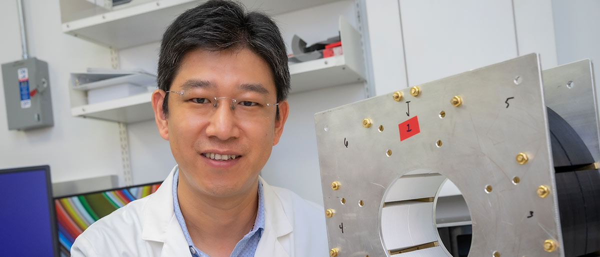
MRI scanner for radiation
Dr. Xun Jia, a Professor of Radiation Oncology, in collaboration with Dr. Anke Henning, Professor of Radiology and Director of the Advanced Imaging Research Center, received a $250,000 grant to develop a new MRI scanner that will help keep radiation focus on tumors in radiotherapy. Instead of using MRI imaging conducted days or weeks ago, the MRI Dr. Jia is developing will attach to radiotherapy equipment and show MRI images immediately before or during radiation treatment. This will help see the tumor and target the radiation. The current state-of-the-art radiotherapy uses cone-beam computed tomography attached to radiotherapy equipment to guide radiation delivery. The new MRI scanner will enable better tumor visualizations without the concern of X-ray exposure in computed tomography.

“We need to see where the tumor is at the moment of treatment before we can fire the radiation to kill the cancer. The new scanner will allow us to see the tumor,” Dr. Jia said. “Although two commercial systems became available lately, they suffer from bulky designs, suboptimal functions for radiation therapy applications, and, most importantly, high developmental costs.”
The MRI’s real-time imaging allows radiation oncologists to account for slight tumor movement and help to keep radiation on the tumor to spare healthy tissue.
“Patient anatomy changes during a treatment course over weeks, such as tumor shape, size, and relative position to nearby organs, as well as during treatment delivery due to, for example, respiratory motion,” Dr. Jia said. “Radiation oncologists need to know the exact size, shape, and position of a tumor at the moment of treatment.”
Drs. Jia and Henning expect the device’s development to take about two years, and they will seek a patent. Dr. Jia said it could be a significant contribution to the fight against cancer because more than half of cancer patients receive radiation treatment.
Dr. Arteaga holds The Lisa K. Simmons Distinguished Chair in Comprehensive Oncology.
Dr. Henning holds the Terry and Robert B. Rowling Chair.
Dr. Singal holds the David Bruton, Jr. Professorship in Clinical Cancer Research and is a Dedman Family Scholar in Clinical Care.
Dr. Zhu holds the Kern Wildenthal, M.D., Ph.D., Distinguished Professorship in Pediatric Research.